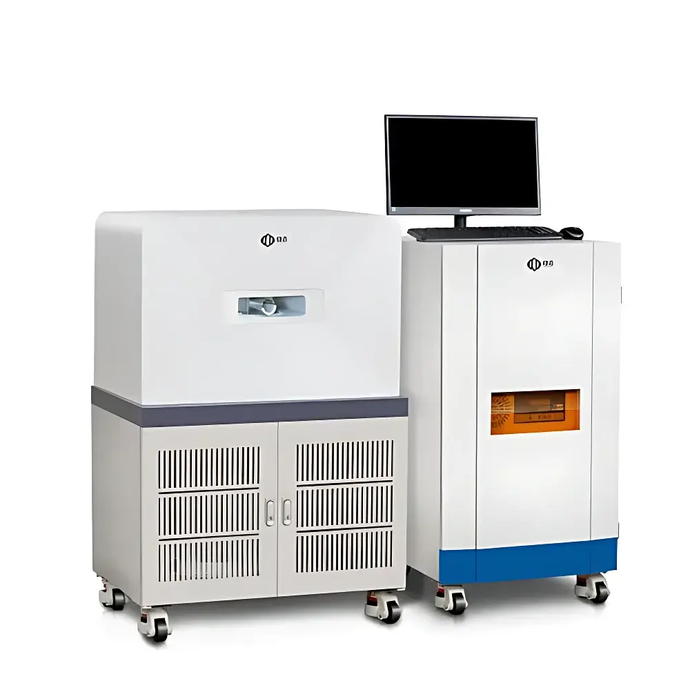
NIUMAG NM21-060H-1 Small Animal MRI System
| Brand | NIUMAG |
|---|---|
| Origin | Jiangsu, China |
| Manufacturer Type | Authorized Distributor |
| Country of Origin | China |
| Model | NM21-060H-1 |
| Pricing | Upon Request |
Overview
The NIUMAG NM21-060H-1 is a compact, benchtop 0.5 Tesla permanent-magnet small animal magnetic resonance imaging (MRI) system engineered for preclinical research in academic laboratories, pharmaceutical R&D centers, and contract research organizations (CROs). Unlike superconducting MRI systems requiring liquid helium cryogenics and RF-shielded rooms, this system employs a high-stability rare-earth permanent magnet—eliminating the need for cryogenic cooling, quench management, or dedicated Faraday cage infrastructure. It operates on standard 220 V AC power with minimal spatial footprint (< 2 m² floor space) and ambient temperature tolerance (18–26 °C), enabling deployment in conventional laboratory environments without structural modifications. The system leverages spin-echo and gradient-echo pulse sequences to generate high-contrast T1-, T2-, and proton density-weighted images of live rodents, supporting longitudinal in vivo studies with non-invasive, radiation-free anatomical and functional assessment.
Key Features
- 0.5 T open-bore permanent magnet configuration with inherent field homogeneity (≤ 10 ppm over 40 mm DSV) and thermal stability (drift < 0.01 ppm/h)
- No cryogens, no RF shielding room required—reduces total cost of ownership and accelerates installation
- Modular RF probe options: interchangeable surface coils optimized for mice (10–30 g) and rats (up to 200 g), with adjustable positioning for reproducible animal centering
- Integrated physiological monitoring interface (optional): supports respiratory gating and ECG synchronization for motion artifact suppression
- Three-step automated imaging workflow: auto-shim → auto-tune → acquisition—minimizing operator dependency and training time
- Real-time parameter adjustment: user-defined TR/TE, flip angle, matrix size (up to 256 × 256), slice thickness (0.3–3.0 mm), and FOV (20–80 mm)
- Low maintenance architecture: solid-state gradient amplifiers, passive shimming, and sealed magnet assembly with > 10-year field stability warranty
Sample Compatibility & Compliance
The NM21-060H-1 accommodates live rodents up to 200 g under isoflurane anesthesia (compatible with optional integrated gas anesthesia delivery module). Its bore diameter (60 mm) and maximum sample height (120 mm) support supine or prone positioning with thermoregulated animal beds (37 °C ± 0.5 °C). The system complies with IEC 61000-6-3 (EMC emissions) and IEC 61000-6-1 (immunity), and meets essential safety requirements per ISO 13485:2016 for medical device-related research instrumentation. While not FDA-cleared as a diagnostic device, it is validated for GLP-compliant preclinical study execution—including audit-ready electronic records when paired with optional 21 CFR Part 11–compliant software modules. All imaging protocols adhere to widely accepted preclinical MRI standards (e.g., ACR–AAPM–SIIM Practice Guidelines for Small Animal MRI).
Software & Data Management
The system ships with two integrated software suites: the NIUMAG MRI Acquisition Suite and the NIUMAG Image Processing Toolkit. The Acquisition Suite provides an intuitive GUI with sequence library management, real-time k-space visualization, and hardware calibration tools. Pulse parameters are fully editable—including RF pulse width, amplitude, and gradient timing—enabling advanced users to implement custom sequences while retaining factory-validated presets for routine T1/T2 mapping, diffusion-weighted imaging (DWI), and MR angiography (MRA). The Image Processing Toolkit supports DICOM import/export, ROI-based quantitative analysis (T1/T2 relaxation time extraction, signal intensity profiling), multi-planar reformatting (MPR), volume rendering, distance/angle measurement, threshold segmentation, and batch-mode pseudo-color mapping. All processed data export to CSV, NIfTI, or MATLAB-compatible formats for downstream statistical analysis in MATLAB, Python (NiBabel), or R.
Applications
- Oncology: Longitudinal tumor volume quantification, necrosis assessment, and contrast-enhanced permeability mapping using Gd-based or iron oxide nanoparticles
- Metabolic disease modeling: Adipose tissue distribution analysis (subcutaneous vs. visceral fat), hepatic steatosis grading, and pancreatic islet morphology tracking
- Neuroscience: Hippocampal volumetry, cortical lesion characterization, and cerebral blood volume (CBV) mapping in stroke or neurodegeneration models
- Nanomedicine: In vivo biodistribution kinetics, carrier degradation monitoring, and target engagement validation via relaxivity-based contrast enhancement
- Cardiovascular research: Cine-MRI for left ventricular ejection fraction (LVEF), wall thickening, and myocardial edema detection
FAQ
Does the NM21-060H-1 require liquid helium or nitrogen?
No. It uses a passively stabilized 0.5 T rare-earth permanent magnet with zero cryogen consumption.
Can the system perform quantitative T1/T2 mapping?
Yes—via built-in multi-echo spin-echo and variable-TR inversion-recovery sequences, with pixel-wise fitting and exportable relaxation maps.
Is RF shielding necessary for operation?
No. The system’s low-field design and optimized gradient shielding enable stable operation in standard lab environments without RF cages.
What anesthesia compatibility does it support?
Full integration with commercial isoflurane vaporizers and nose-cone delivery systems; optional temperature- and respiration-monitored animal bed available.
How is data security and regulatory compliance addressed?
Optional 21 CFR Part 11 add-on enables electronic signatures, audit trails, role-based access control, and secure DICOM storage—supporting FDA-regulated preclinical submissions.
")