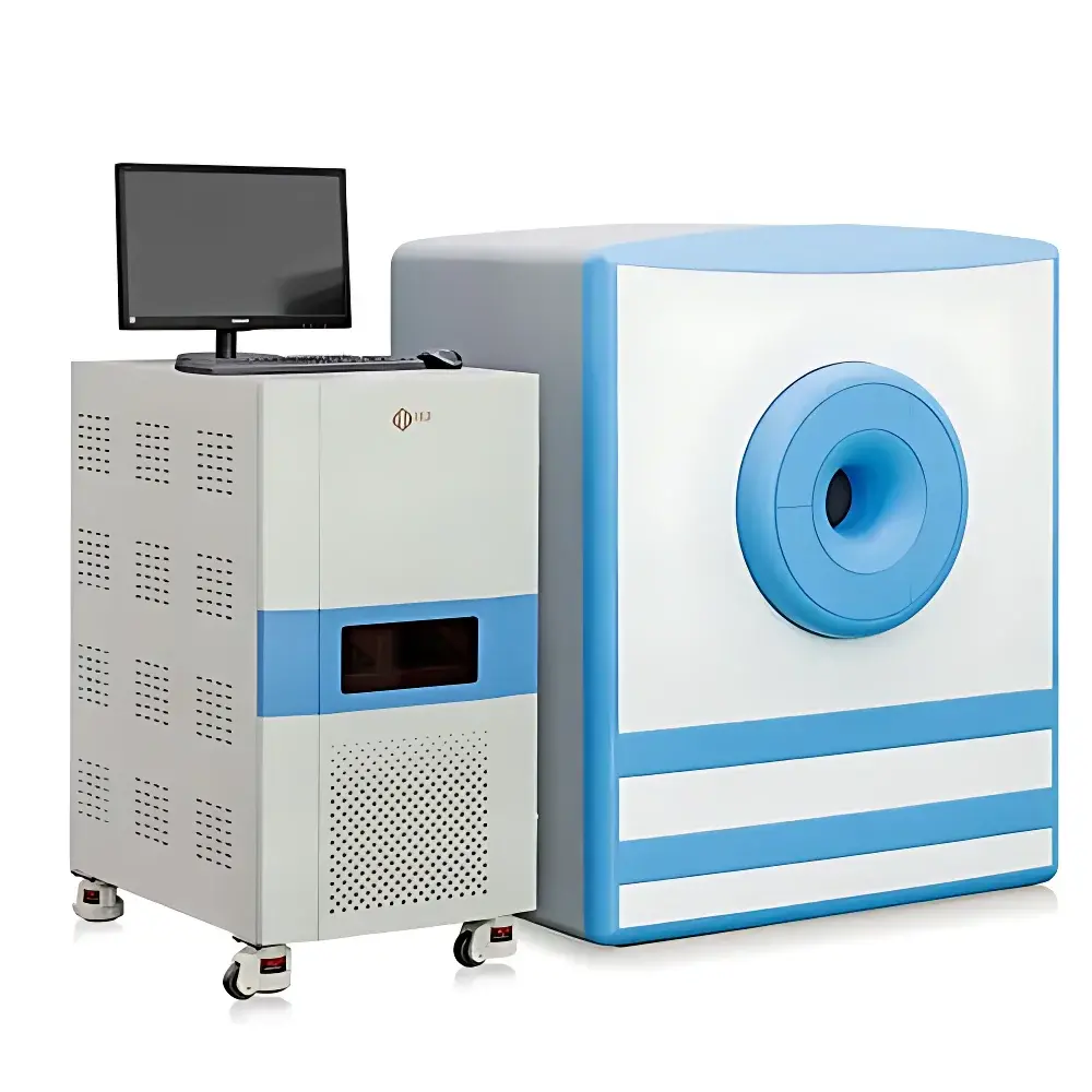
NIUMAG NM42-040H-I 1.0 T Small Animal Magnetic Resonance Imaging System
| Brand | NIUMAG |
|---|---|
| Origin | Shanghai, China |
| Manufacturer Type | Authorized Distributor |
| Country of Origin | China |
| Model | NM42-040H-I |
| Instrument Type | Magnetic Resonance Imaging (MRI) System |
| Magnetic Field Strength | 1.0 ± 0.08 T |
| Magnet Type | Permanent Magnet |
| Field Homogeneity | ≤20 ppm over Ø40 mm × H40 mm DSV |
| Imaging FOV | Ø40 mm × 40 mm (height) |
| Minimum Slice Thickness | 0.8 mm |
| Spatial Resolution (in-plane) | 0.08 mm |
| Gradient System | High-Performance Integrated Gradient Set |
| RF Coil Configuration | Animal-Specific Tunable RF Coils |
Overview
The NIUMAG NM42-040H-I is a benchtop 1.0 Tesla permanent-magnet small animal MRI system engineered for preclinical in vivo imaging research. Operating on the fundamental principles of nuclear magnetic resonance—specifically the detection and spatial encoding of proton (¹H) signal relaxation dynamics—the system delivers high-fidelity T₁-, T₂-, and proton density-weighted contrast without ionizing radiation. Its 1.0 T field strength, among the highest available in compact permanent-magnet MRI platforms, enables enhanced signal-to-noise ratio (SNR), improved spectral separation, and greater sensitivity to subtle tissue contrast changes compared to lower-field systems (e.g., 0.3–0.5 T). The magnet’s homogeneity—≤20 ppm over a 40 mm spherical volume—ensures robust image fidelity across the full imaging volume, supporting quantitative longitudinal studies in rodent models under standardized acquisition protocols.
Key Features
- 1.0 ± 0.08 T permanent magnet with active shimming and temperature-stabilized design for long-term field stability and reproducibility
- High-performance gradient subsystem delivering precise spatial encoding for multiplanar (axial, coronal, sagittal) and oblique acquisitions
- Animal-specific RF coil suite—including tunable surface and volume coils—optimized for mice (≥15 g) and rats (up to 500 g), enabling impedance matching and SNR maximization per subject
- Sub-millimeter imaging capability: minimum slice thickness of 0.8 mm with isotropic in-plane resolution down to 0.08 mm
- Three-step intuitive acquisition workflow (Setup → Scan → Analyze), preserving NIUMAG’s legacy usability while integrating modern pulse sequence flexibility
- Compact footprint (≤1.2 m² floor space) and low power consumption (<3 kW), suitable for dedicated lab environments without cryogen or RF-shielded room requirements
Sample Compatibility & Compliance
The NM42-040H-I supports non-invasive longitudinal imaging of live rodents within its defined imaging volume (Ø40 mm × 40 mm height), accommodating restrained or anesthetized subjects via compatible stereotactic holders and physiological monitoring interfaces (respiratory gating, temperature control). All hardware and software components comply with IEC 61000-6-3 (EMC emissions) and IEC 61000-6-2 (immunity) standards. Data acquisition workflows are structured to support Good Laboratory Practice (GLP) documentation requirements, including user-access logging, parameter versioning, and DICOM-compliant metadata embedding. While not FDA-cleared for clinical use, the system adheres to ASTM F2503-21 guidelines for MRI safety labeling and supports audit-ready operation for academic and pharmaceutical preclinical facilities.
Software & Data Management
The system runs NIUMAG’s proprietary MR Console v5.x platform, featuring a modular architecture that separates acquisition control, reconstruction, and post-processing modules. Pulse sequences include spin echo (SE), fast spin echo (FSE), gradient echo (GRE), inversion recovery (IR), and diffusion-weighted imaging (DWI)—all fully customizable via editable sequence parameters. Reconstruction employs parallel imaging acceleration (SENSE-based) and zero-filled Fourier interpolation. Processed data exports natively to DICOM 3.0 format and supports NIfTI conversion for integration with FSL, SPM, or AFNI. The integrated image analysis suite provides semi-automated ROI segmentation, volumetric quantification (e.g., tumor volume, fat fraction), T₁/T₂ mapping, and time-series intensity profiling—all traceable with embedded timestamps, operator IDs, and acquisition parameter hashes for regulatory traceability.
Applications
- Oncology: Longitudinal monitoring of orthotopic and subcutaneous tumor growth, response to chemotherapeutics or immunotherapies, and evaluation of nanocarrier biodistribution
- Metabolic disease modeling: Quantitative assessment of adipose tissue distribution (subcutaneous vs. visceral), hepatic steatosis, and pancreatic morphology in diet-induced obesity and type 2 diabetes models
- Neuroscience: Structural brain phenotyping, stroke lesion volumetry, and white matter integrity assessment using diffusion tensor imaging (DTI)
- Contrast agent development: In vivo pharmacokinetic profiling of T₁- and T₂-based agents, relaxivity validation, and target engagement verification via dynamic contrast-enhanced (DCE) MRI
- Cardiovascular research: Cine-MRI for cardiac function metrics (ejection fraction, wall thickening), myocardial edema detection, and infarct sizing
- Toxicology & safety pharmacology: Detection of organ-level morphological alterations (e.g., renal cysts, hepatic necrosis) following compound exposure
FAQ
Is the NM42-040H-I compliant with FDA 21 CFR Part 11 for electronic records and signatures?
The system supports audit trail generation, user authentication, and parameter locking for critical acquisition steps; however, full Part 11 compliance requires site-specific validation and procedural documentation—not inherent to the instrument alone.
Can the system perform functional MRI (fMRI) or spectroscopy (MRS)?
While optimized for structural and quantitative MRI, the platform does not currently support BOLD fMRI or localized MRS due to gradient slew rate and RF bandwidth limitations inherent to permanent-magnet architecture.
What anesthesia delivery options are supported?
The system is compatible with standard rodent vaporizer-based isoflurane systems (e.g., Euthanex, SomnoSuite) via external gas line routing through the scanner bore interface port.
Is remote operation or telemonitoring possible?
Yes—acquisition can be initiated and monitored remotely via secure VNC or RDP connection to the host workstation, provided local network policies permit encrypted traffic.
Does NIUMAG provide application support for protocol optimization?
NIUMAG offers tiered technical support packages, including on-site protocol development assistance for common disease models, sequence customization consulting, and DICOM archive integration guidance.


