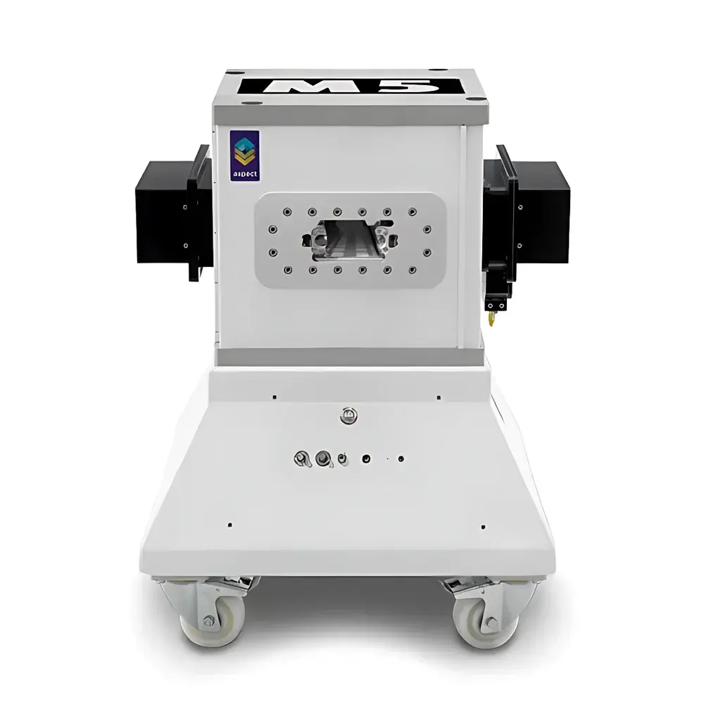
NIUMAG Aspect M5 Small Animal MRI System
| Brand | NIUMAG |
|---|---|
| Origin | Israel |
| Manufacturer Type | Authorized Distributor |
| Origin Category | Imported |
| Model | M5 |
| Instrument Type | Nuclear Magnetic Resonance Imaging (MRI) System |
| Magnet Type | Permanent Magnet, 1 Tesla |
| Weight Capacity | Up to 700 g (mouse/rat) |
| Imaging Modes | 2D/3D, In Vivo, Ex Vivo, In Vitro |
| Cooling | Passive (No Cryogens or Chiller Required) |
| Shielding | Self-Shielded (Zero Stray Field) |
| Typical Resolution | ≤ 234 µm (T2-weighted), ≤ 195 µm (T1-weighted) |
| Acquisition Time | ~4.8–9.1 min per scan |
| Compliance | Designed for ISO 13485-aligned lab environments |
Overview
The NIUMAG Aspect M5 Small Animal MRI System is a compact, self-shielded, permanent-magnet-based nuclear magnetic resonance imaging platform engineered for high-fidelity in vivo and ex vivo phenotypic imaging of rodents up to 700 g. Operating at a stable 1 Tesla field strength, the system leverages robust permanent magnet architecture—eliminating cryogenic cooling, RF shielding rooms, and external infrastructure dependencies. Its fully integrated design enables installation directly behind biosafety containment barriers (e.g., Class II BSCs or isolators), supporting longitudinal studies under strict biocontainment protocols without compromising image fidelity or operator safety. The Aspect M5 employs standard spin-echo (SE) and fast spin-echo (FSE) pulse sequences, delivering quantitative T1- and T2-weighted contrast with sub-200 µm in-plane resolution under default acquisition parameters—making it suitable for structural, functional, and pharmacokinetic investigations in preclinical research.
Key Features
- Self-shielded 1 T permanent magnet: Zero stray magnetic field (< 0.5 mT at 1 m distance), enabling safe placement in shared laboratory spaces and adjacent to sensitive instrumentation.
- No cryogen dependency: Passive thermal stabilization eliminates liquid helium consumption, chiller units, or scheduled quench maintenance—reducing total cost of ownership and operational downtime.
- One-touch acquisition workflow: Intuitive touchscreen interface with preconfigured protocols for common applications (e.g., brain anatomy, tumor volume quantification, cardiac cine, abdominal fat mapping).
- Biosafety-integrated deployment: Compact footprint (W × D × H ≈ 120 × 90 × 110 cm) allows positioning within ISO Class 5–7 cleanrooms or behind laminar flow hoods without airflow disruption.
- Multi-contrast capability: Native support for T1-, T2-, PD-, and diffusion-weighted imaging via adjustable TR/TE, ETL, NEX, FOV, and matrix parameters.
- Modular RF coil ecosystem: Interchangeable volume and surface coils optimized for head, thorax, abdomen, and extremity imaging—each calibrated for SNR optimization at 42.6 MHz (¹H Larmor frequency at 1 T).
Sample Compatibility & Compliance
The Aspect M5 accommodates live mice and rats (up to 700 g) under isoflurane anesthesia, as well as excised tissues, organoids, and tissue phantoms. Integrated physiological monitoring (respiratory gating, temperature control via heated air system) ensures motion artifact suppression during long acquisitions. All hardware and software components comply with IEC 61000-6-3 (EMC emissions) and IEC 61000-6-1 (immunity) standards. While not a medical device, the system’s architecture supports audit-ready data handling—raw k-space and DICOM-compliant image exports include timestamped metadata, user ID, sequence parameters, and calibration logs—facilitating adherence to GLP documentation requirements and internal SOP validation.
Software & Data Management
The proprietary Aspect MRI Suite provides full sequence programming (Pulse Sequence Development Environment), real-time reconstruction, and post-processing tools including region-of-interest (ROI) analysis, volumetric segmentation, and intensity normalization. Image data are exported in DICOM 3.0 format (conforming to PS3.10) and raw binary formats for third-party analysis (e.g., MATLAB, FSL, ITK-SNAP). Audit trails record all parameter modifications, user logins, and export events—meeting traceability expectations under FDA 21 CFR Part 11 when deployed with institutional electronic signature policies. Data storage is managed via encrypted local SSD or network-attached storage (NAS) with configurable retention policies.
Applications
- Oncology: Longitudinal tracking of orthotopic/subcutaneous tumor growth, metastatic burden, and treatment response (e.g., anti-angiogenic therapy, immunotherapy).
- Neuroscience: Hippocampal volumetry, cortical lesion mapping, white matter integrity assessment (via T2 relaxometry), and functional connectivity surrogate metrics.
- Cardiovascular research: Cine-MRI for ejection fraction, wall motion analysis, and myocardial edema quantification post-ischemia.
- Metabolic disease: Visceral vs. subcutaneous adipose tissue differentiation, hepatic steatosis grading, and pancreatic islet mass estimation.
- Developmental biology: Embryonic organogenesis imaging (ex vivo), placental perfusion modeling, and fetal brain maturation studies.
- Contrast agent development: Pharmacokinetic profiling of Gd-based, iron oxide, or CEST agents—including relaxivity quantification and biodistribution kinetics.
FAQ
Does the Aspect M5 require a dedicated RF-shielded room?
No. Its fully self-shielded permanent magnet generates negligible external fringe field, permitting installation in standard laboratory environments without Faraday cage construction.
Can the system be used for longitudinal studies across multiple time points?
Yes. The passive magnet stability ensures consistent field homogeneity over years, and built-in respiratory gating + temperature regulation support repeatable, low-variability imaging sessions.
Is DICOM export compliant with PACS integration?
Yes. All reconstructed images are exported in DICOM 3.0 format with modality-specific SOP classes (MR Image Storage), supporting direct ingestion into institutional PACS or preclinical imaging archives.
What coil options are available for specialized anatomical regions?
NIUMAG offers a suite of validated coils: 35 mm transmit/receive volume coil (head), 70 mm volume coil (whole-body), and 25 mm surface coil (spine, hindlimb); all include automatic tuning/matching and integrated preamplifiers.
How is system performance verified and maintained?
Daily QA includes phantom-based SNR, geometric distortion, and center frequency checks using the included NIST-traceable test object; annual calibration is optional and performed remotely via secure diagnostic port.


