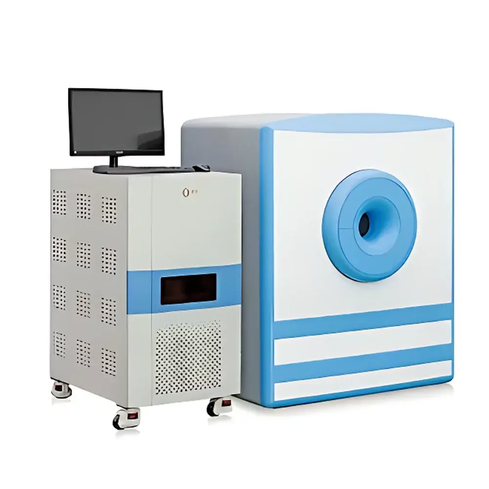
NIUMAG NM42 Small Animal In Vivo MRI System
| Brand | NIUMAG |
|---|---|
| Origin | Jiangsu, China |
| Manufacturer Type | Authorized Distributor |
| Country of Origin | China |
| Model | NM42 |
| Instrument Type | 1.0 Tesla Permanent-Magnet Benchtop MRI System |
| Animal Weight Range | 1–45 g |
| Maximum Sample Width | < 40 mm |
| Minimum Sample Volume | ≥ 100 µL |
| Minimum Slice Thickness | 0.8 mm |
| Field Homogeneity | Optimized for high SNR and spatial resolution |
| Anesthesia Compatibility | Optional integrated gas anesthesia module |
| Shielding Requirement | None — no RF-shielded room required |
Overview
The NIUMAG NM42 is a compact, 1.0 Tesla permanent-magnet benchtop magnetic resonance imaging (MRI) system engineered specifically for preclinical in vivo imaging of small rodents and ex vivo analysis of biological solutions. Unlike superconducting MRI systems requiring cryogens and RF-shielded facilities, the NM42 leverages a high-stability permanent magnet architecture to deliver robust T₁- and T₂-weighted imaging with clinically relevant contrast mechanisms—without ionizing radiation, surgical intervention, or physiological stress. Its core measurement principle is based on nuclear magnetic resonance signal detection from hydrogen protons (¹H) in water and lipid environments, enabling non-invasive, longitudinal monitoring of anatomical structure, tissue perfusion, lesion development, and contrast agent biodistribution. Designed for laboratories with limited infrastructure, the NM42 operates in standard ambient environments and supports rapid acquisition protocols—typical scan times range from 2 to 8 minutes depending on resolution and sequence selection.
Key Features
- 1.0 T permanent magnet with optimized field homogeneity—enabling high signal-to-noise ratio (SNR), sub-millimeter spatial resolution (down to 0.8 mm slice thickness), and multiplanar reformatting (axial, sagittal, coronal, oblique) without hardware repositioning
- Non-invasive, radiation-free imaging—ideal for longitudinal studies across multiple timepoints in the same animal, minimizing inter-subject variability and ethical burden
- Wide subject compatibility—accommodates mice, rats, and other small mammals up to 45 g body weight and ≤40 mm transverse width
- Three-step automated workflow—integrated auto-shim, auto-tune, and auto-centering routines eliminate dependence on operator expertise in NMR physics
- Modular expandability—including optional precision gas anesthesia delivery (isoflurane/O₂), physiological monitoring interfaces (respiratory gating, temperature control), and dedicated RF coils for head/body applications
- Low total cost of ownership—no liquid helium, no RF shielding, no specialized HVAC or power conditioning; routine maintenance limited to coil cleaning and software updates
Sample Compatibility & Compliance
The NM42 supports both in vivo and ex vivo sample configurations. In vivo applications include tumor xenograft monitoring, pharmacokinetic evaluation of contrast agents, therapeutic response assessment (e.g., anti-angiogenic or cytostatic drug efficacy), and neuroanatomical phenotyping. Ex vivo compatibility extends to quantitative relaxometry of nanoparticle suspensions, ionic solutions, microbial cultures, and tissue homogenates (minimum volume: 100 µL). All imaging protocols comply with standard preclinical MRI reporting guidelines (ARRIVE 2.0) and support traceable data acquisition under GLP-aligned workflows. While not FDA-cleared for human use, the system adheres to IEC 62353 (electrical safety) and ISO 13485-aligned quality management practices in manufacturing and distribution. Data integrity is preserved through timestamped DICOM export and audit-ready metadata logging.
Software & Data Management
The NM42 is operated via two tightly integrated software suites: the NIUMAG MRI Acquisition Platform and the NIUMAG Image Processing Suite. The acquisition platform provides access to spin-echo, gradient-echo, fast spin-echo, and inversion-recovery pulse sequences—with fully adjustable TR/TE/TI, flip angle, bandwidth, and matrix size. Pulse parameter modulation is implemented via intuitive sliders and preset templates, eliminating the need for command-line input or pulse sequence coding. All acquisitions are stored in DICOM 3.0 format with embedded scanner parameters and calibration logs. The image processing suite enables ROI-based quantification (T₁/T₂ mapping, signal intensity normalization), pseudo-color overlay, 3D volume rendering, distance/angle measurement, threshold segmentation, and batch-mode data export (CSV, NIfTI, TIFF). Both modules support 21 CFR Part 11-compliant user authentication, electronic signatures, and audit trail generation when deployed on validated Windows Server environments.
Applications
- Preclinical oncology: Detection and volumetric tracking of subcutaneous or orthotopic tumors; evaluation of necrosis, edema, and vascular permeability using dynamic contrast-enhanced (DCE) MRI
- Contrast agent development: Quantitative r₁/r₂ relaxivity measurement in vitro and in vivo; biodistribution kinetics and clearance profiling
- Neuroscience research: High-resolution structural imaging of mouse brain anatomy; lesion modeling in stroke or demyelination studies
- Nanomedicine characterization: Relaxometric fingerprinting of iron oxide nanoparticles, manganese-based probes, or gadolinium chelates in solution phase
- Microbiology & biophysics: Assessment of cellular aggregation, biofilm formation, or ion-induced T₂ shortening in suspension cultures
FAQ
Does the NM42 require an RF-shielded room?
No. The permanent magnet design and low-field architecture eliminate the need for external RF shielding—installation is possible in standard laboratory spaces with standard electrical outlets.
Can the system perform quantitative T₁ and T₂ mapping?
Yes. With appropriate inversion-recovery or multi-echo spin-echo protocols, the NM42 supports pixel-wise T₁ and T₂ calculation using built-in fitting algorithms and ROI-based statistical analysis.
Is anesthesia integration mandatory for in vivo imaging?
Anesthesia is strongly recommended for motion artifact suppression during in vivo scans but is not hardware-mandatory—the system accepts third-party anesthesia units via analog/digital I/O ports.
What file formats are supported for data export?
DICOM 3.0 (primary), NIfTI 1.1, TIFF (8/16-bit), CSV (ROI statistics), and MATLAB (.mat) for advanced post-processing.
How is system calibration maintained over time?
The NM42 performs daily auto-shim and reference-scan calibration; long-term stability is verified annually using standardized phantom measurements (e.g., ACR MRI Phantom) per ASTM D7291 guidelines.


