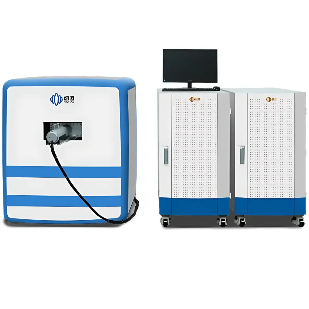
NIUMAG NM42-060H-I-1 Small Animal In Vivo MRI System
| Brand | NIUMAG |
|---|---|
| Origin | Jiangsu, China |
| Manufacturer Type | Authorized Distributor |
| Country of Origin | China |
| Model | NM42-060H-I-1 |
| Instrument Type | Low-Field Nuclear Magnetic Resonance (LF-NMR) Imaging System |
| Magnet Type | Permanent Magnet |
| Field Strength | 1.0 T ± 0.05 T |
| Homogeneity | ≤30 ppm over 60 mm DSV |
| Sample Capacity | 1–350 g (mice, rats, excised tissues) |
| Imaging Modes | T1-weighted, T2-weighted, Proton Density-weighted, Water-Fat Suppressed Imaging |
Overview
The NIUMAG NM42-060H-I-1 Small Animal In Vivo MRI System is a dedicated low-field nuclear magnetic resonance (LF-NMR) imaging platform engineered for longitudinal, non-invasive structural and functional assessment in preclinical rodent models of neurological injury. Operating at a stable 1.0 Tesla field strength generated by a high-homogeneity permanent magnet, the system leverages the intrinsic contrast mechanisms of proton relaxation (T1, T2, PD) to resolve anatomical boundaries, lesion volume, edema progression, hemorrhage evolution, and tissue viability without ionizing radiation or surgical intervention. Unlike high-field MRI systems requiring cryogenic cooling and RF-shielded rooms, this instrument employs a self-shielded, air-cooled permanent magnet architecture—enabling installation in standard laboratory environments with minimal infrastructure requirements. Its design prioritizes reproducibility, quantitative stability, and compliance with GLP-aligned workflows for translational neuroscience research.
Key Features
- Permanent magnet configuration: Eliminates dependency on liquid helium, nitrogen, or external chiller units—reducing operational cost and footprint.
- High spatial resolution: Achieves sub-100 µm in-plane resolution under optimized acquisition protocols, sufficient for delineating cortical layers, hippocampal subfields, ventricular margins, and lesion cores in murine brains.
- Multi-contrast capability: Supports T1-weighted, T2-weighted, proton density-weighted, and water-fat suppressed (e.g., Dixon-based) acquisitions—enabling differential characterization of vasogenic edema, cytotoxic swelling, gliosis, and demyelination.
- Integrated animal handling system: Includes temperature-regulated physiological monitoring (respiratory gating, rectal thermistor), stereotactic head fixation, and gas anesthesia delivery (isoflurane/O₂) compatible with chronic imaging sessions.
- Robust gradient performance: 25 G/cm maximum gradient strength with <100 µs rise time supports diffusion-weighted imaging (DWI) and basic perfusion-sensitive sequences for acute ischemia modeling.
Sample Compatibility & Compliance
The NM42-060H-I-1 accommodates live mice (15–35 g), rats (200–350 g), and excised brain tissues within a 60 mm diameter spherical volume (DSV). It is validated for use in standardized traumatic brain injury (TBI) models—including controlled cortical impact (CCI), fluid percussion injury (FPI), and weight-drop; focal cerebral ischemia models (MCAO); LPS-induced neuroinflammation; and hypoxic-ischemic encephalopathy (HIE) paradigms. All hardware and software components conform to IEC 61000-6-3 (EMC emission) and IEC 61000-6-2 (immunity) standards. Data acquisition and storage support audit-trail functionality aligned with FDA 21 CFR Part 11 requirements when deployed with optional electronic signature modules. The system meets ISO 13485 design control principles for Class IIa medical device development tools used in preclinical safety and efficacy evaluation.
Software & Data Management
Acquisition and reconstruction are managed via NIUMAG’s proprietary NMI-Studio v4.x platform, featuring DICOM 3.0 export, BIDS-compliant metadata tagging, and native support for NIfTI format. The software includes automated brain extraction (BET), co-registration to Allen Mouse Brain Atlas (v3), and voxel-wise quantification of T1/T2 relaxation times using monoexponential fitting. Raw k-space data is preserved for offline processing in MATLAB, Python (NiBabel, DIPY), or FSL. Audit logs record operator ID, sequence parameters, acquisition timestamps, and calibration events—essential for regulatory submissions and multi-site study harmonization. Optional integration with LabArchives ELN enables direct embedding of reconstructed images and ROI statistics into electronic lab notebooks.
Applications
- Longitudinal monitoring of lesion evolution in murine TBI models: Quantifying contusion volume, perilesional edema kinetics, and white matter tract integrity via DTI-derived FA maps.
- Evaluation of neuroprotective agents in MCAO: Assessing infarct size reduction, blood-brain barrier permeability changes (via dynamic contrast-enhanced MRI), and reperfusion efficacy.
- Tracking magnetic nanoparticle biodistribution: Monitoring accumulation in inflamed brain parenchyma post-systemic injection using T2* susceptibility mapping.
- Pharmacokinetic-pharmacodynamic (PK-PD) correlation: Linking regional gadolinium-enhancement patterns with histopathological markers of angiogenesis or macrophage infiltration.
- Validation of optical imaging findings: Cross-modal registration of MRI-derived anatomical priors with two-photon or bioluminescence datasets.
FAQ
Is this system suitable for functional MRI (fMRI) studies in awake mice?
No—the NM42-060H-I-1 is optimized for structural and relaxation-based contrast imaging. It does not support echo-planar imaging (EPI) or real-time physiological noise correction required for BOLD fMRI in unanesthetized subjects.
Can the system perform quantitative T1/T2 mapping?
Yes—built-in inversion recovery (IR) and multi-echo spin-echo (MESE) sequences enable pixel-wise T1 and T2 relaxation time estimation with coefficient of variation <5% across repeated scans.
What level of technical support and service coverage is provided?
NIUMAG offers on-site installation, operator training, and 24-month comprehensive warranty including magnet stability certification, gradient coil recalibration, and software updates. Extended service contracts include annual homogeneity verification and RF coil performance validation.
Does the system comply with GLP documentation requirements for IND-enabling studies?
Yes—when configured with optional 21 CFR Part 11 add-ons (user access controls, electronic signatures, immutable audit trails), the system supports GLP-compliant data generation for regulatory submissions to FDA, EMA, and PMDA.
Are custom pulse sequences available for advanced contrast mechanisms?
Third-party sequence development is supported via C++ SDK and MATLAB interface; users may implement custom diffusion tensor, magnetization transfer, or chemical exchange saturation transfer (CEST) protocols subject to gradient and SAR limitations.


