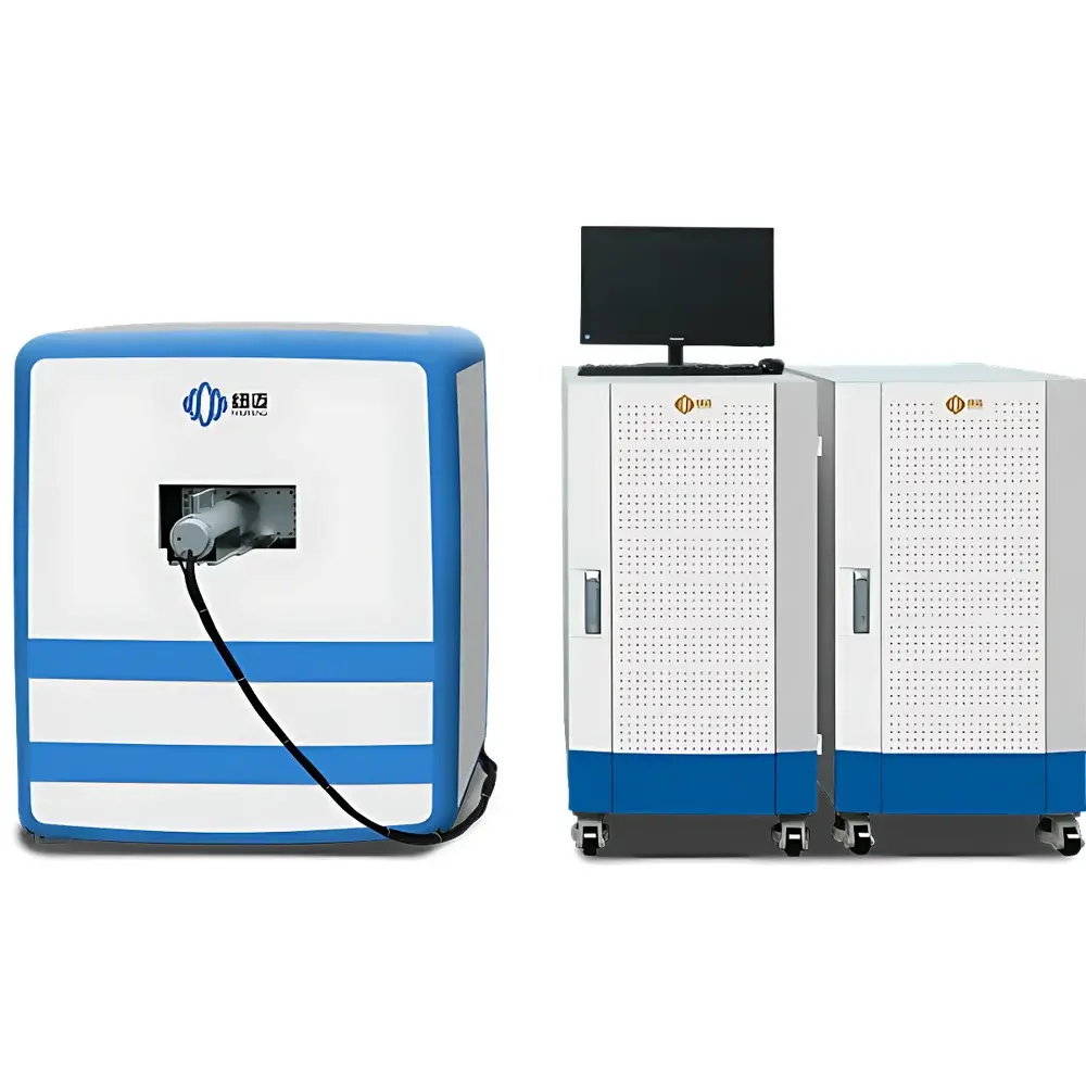
NIUMAG NM42-060H-I Small Animal MRI System
| Brand | NIUMAG |
|---|---|
| Origin | Jiangsu, China |
| Manufacturer Type | Authorized Distributor |
| Country of Origin | China |
| Model | NM42-060H-I |
| Instrument Type | Low-Field Nuclear Magnetic Resonance Imaging System |
| Sample Type | Solid-Liquid Dual-Mode (In Vivo Mice, Ex Vivo Tissues, Contrast Agents, Magnetic Nanoparticles) |
| Magnet Type | Permanent Magnet |
| Static Field Strength | 1.0 T ± 0.05 T |
| Magnet Homogeneity | ≤30 ppm over Ø60 mm DSV |
| Imaging Matrix Support | Up to 512 × 512 × 128 |
| RF Coil Options | φ25 mm / φ30 mm / φ35 mm / φ45 mm / φ60 mm Dedicated Animal Body Coils |
| Integrated Anesthesia & Respiratory Monitoring | Yes |
| Temperature-Controlled Animal Bed | Yes |
| Pulse Sequence Flexibility | Graphical GUI + Python Scripting Support |
| Data Throughput Architecture | Gigabit Ethernet-Based Real-Time Acquisition Platform |
| Bit Error Rate | One Order of Magnitude Lower Than Conventional Open-Source Spectrometers |
Overview
The NIUMAG NM42-060H-I Small Animal MRI System is a purpose-built, low-field nuclear magnetic resonance imaging platform engineered for preclinical in vivo studies in murine models. Operating at a stable static magnetic field of 1.0 T ± 0.05 T generated by a high-stability permanent magnet assembly, the system delivers robust signal-to-noise ratio (SNR) and spatial fidelity without reliance on cryogenic cooling or RF-shielded rooms. Its core architecture integrates a proprietary gigabit-speed spectrometer with optimized pulse sequence execution, real-time data streaming, and deterministic signal processing pathways—enabling reproducible acquisition of T1-weighted, T2-weighted, proton density-weighted, and fat-suppressed contrast images. Designed specifically for longitudinal studies in oncology, neurodegeneration, cardiovascular disease, and nanotherapeutic delivery, the NM42-060H-I supports rigorous GLP-aligned experimental workflows through hardware-level respiratory gating, physiological monitoring, and thermal stabilization of live subjects during scanning.
Key Features
- Permanent magnet design eliminates liquid helium consumption, nitrogen boil-off, and structural shielding requirements—reducing total cost of ownership and enabling installation in standard laboratory environments.
- Integrated gas anesthesia delivery and real-time respiratory monitoring module ensures precise control of inhalational anesthetics (e.g., isoflurane) and synchronization of image acquisition with respiratory cycles via hardware-triggered gating.
- Dedicated animal handling platform includes active temperature regulation (37 °C ± 0.5 °C) across the full scan duration, minimizing physiological drift and enhancing inter-scan reproducibility.
- Multi-diameter RF body coils (Ø25–Ø60 mm) support automatic tuning and matching, enabling rapid coil switching and optimal B1 homogeneity for diverse specimen masses (1–350 g).
- Graphical pulse sequence editor with Python API allows full customization of RF excitation, gradient timing, k-space trajectory, and reconstruction logic—facilitating implementation of advanced sequences including RARE, FLASH, EPI, and diffusion-weighted protocols.
- High-fidelity 3D reconstruction up to 512 × 512 × 128 matrix resolution enables sub-millimeter isotropic voxel rendering for volumetric analysis of anatomical structures and lesion progression.
Sample Compatibility & Compliance
The NM42-060H-I accommodates both in vivo murine subjects (mice and rats) and ex vivo tissues, as well as magnetic nanoparticles, MR contrast agents (Gd-based, iron oxide), and hydrogel-based phantoms. Its open-bore configuration and modular coil system permit flexible positioning—including prone, supine, and lateral orientations—without mechanical restraints compromising physiological stability. The system complies with IEC 61000-6-3 (EMC emission standards) and meets essential safety requirements per ISO 13485:2016 for medical device-related research instrumentation. While not intended for human diagnostic use, its acquisition parameters and DICOM export capabilities align with preclinical imaging standards referenced in NIH-funded animal studies and FDA-guided IND-enabling toxicology assessments.
Software & Data Management
Acquisition and reconstruction are managed through NIUMAG’s proprietary NMI-Studio software suite, compliant with DICOM 3.0 Part 10 file structure for seamless integration into PACS and third-party analysis platforms (e.g., ITK-SNAP, FSL, MATLAB). All raw k-space data and reconstructed NIfTI volumes include embedded metadata: scanner parameters, subject ID, anesthesia settings, coil configuration, and timestamped physiological logs. Audit trails record user login sessions, sequence modifications, and export events—supporting 21 CFR Part 11 readiness when deployed under controlled QA/QC frameworks. Data encryption at rest and TLS 1.2–secured remote access options are available for multi-site collaborative studies requiring HIPAA- or GDPR-aligned data governance.
Applications
- Tumor xenograft monitoring: Quantitative assessment of volume change, necrosis fraction, and vascular permeability using dynamic contrast-enhanced (DCE) MRI.
- Neuroimaging: High-resolution structural mapping of hippocampal atrophy, white matter integrity via diffusion tensor imaging (DTI), and functional connectivity in transgenic Alzheimer’s models.
- Nanoparticle biodistribution: Tracking superparamagnetic iron oxide nanoparticle (SPION) accumulation in liver, spleen, and tumor microenvironments using T2* relaxometry.
- Cardiac phenotyping: Cine-MRI for left ventricular ejection fraction, wall thickness dynamics, and myocardial strain analysis in pressure-overload murine models.
- Pharmacokinetic evaluation: Time-resolved contrast agent kinetics for assessing blood-brain barrier integrity and drug delivery efficiency.
FAQ
Is this system suitable for longitudinal studies across multiple timepoints?
Yes—the integrated thermal regulation, non-invasive anesthesia interface, and low SAR RF design enable repeated imaging sessions over days or weeks without compromising animal welfare or data consistency.
Can I import custom pulse sequences developed in MATLAB or Pulseq format?
The system supports Pulseq-compatible sequence definitions and provides Python bindings for direct integration with external simulation and optimization toolchains.
Does the platform support quantitative mapping (T1, T2, ADC)?
Yes—standard inversion-recovery and spin-echo multi-echo protocols are preconfigured; users may extend these using the graphical sequence builder or script-based parameter sweeps.
What level of technical support is provided for method development?
NIUMAG offers application engineering support—including sequence optimization, phantom validation, and protocol documentation—for academic and industrial clients under annual service agreements.
Are DICOM archives compatible with PACS systems used in core imaging facilities?
All exported DICOM series conform to Supplement 119 (Enhanced MR Image Storage) and include required modality-specific attributes for automated ingestion into enterprise PACS environments.