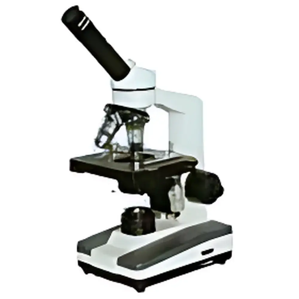
XSP-10CA Trinocular Biological Microscope
| Origin | Shanghai, China |
|---|---|
| Manufacturer Type | Authorized Distributor |
| Origin Category | Domestic (PRC) |
| Model | XSP-10CA |
| Price | USD 520 (FOB Shanghai) |
| Tube | Trinocular (20% beam split for camera port) |
| Condenser | Abbe condenser with adjustable iris diaphragm and filter holder |
| Eyepieces | Wide-field WF10× (18 mm FOV), Huygens H16× (11 mm FOV) |
| Objectives | Achromatic 4×, 10×, 40× (spring-loaded, semi-plan), 100× (oil immersion, spring-loaded, semi-plan) |
| Illumination | Adjustable 3W LED or 6V/30W halogen lamp |
| Power Supply | AC 100–240 V, 50/60 Hz, integrated switching power supply |
Overview
The XSP-10CA Trinocular Biological Microscope is an entry-to-mid-tier upright compound microscope engineered for routine histological examination, educational laboratory instruction, and basic live-cell observation in academic and clinical support settings. It operates on standard brightfield optical principles using Köhler illumination geometry, with an achromatic objective suite corrected for chromatic aberration at two wavelengths (typically 486 nm and 656 nm) and spherical aberration at the central paraxial zone. Its trinocular head features a fixed 80:20 beam splitter ratio—80% directed to the eyepieces for visual observation and 20% transmitted to a C-mount port—enabling simultaneous human viewing and digital image acquisition without optical realignment. Designed for stability and ergonomic operation, the instrument incorporates a rigid cast-aluminum stand, coaxial coarse/fine focusing mechanism with 2 µm minimum graduation, and stage controls with low-friction ball-bearing translation.
Key Features
- Rigid mechanical design with vibration-damped base and precision-machined dovetail-mounted stage for consistent focus retention during extended use.
- Abbe condenser (NA 1.25) with centerable iris diaphragm and integrated filter holder accommodating standard 32 mm round filters (e.g., daylight blue, green interference, ND).
- Dual illumination options: energy-efficient 3W white LED (50,000-hour lifetime, CCT ~5500 K, flicker-free PWM dimming) or replaceable 6V/30W halogen bulb with built-in voltage regulation and heat-absorbing glass.
- Achromatic objective set optimized for parfocality and parcentricity; all objectives feature spring-loaded front lenses and standardized RMS threading (19 mm pitch, 0.706 mm thread depth) for interchangeability with legacy equipment.
- Trinocular head with ±5° interpupillary adjustment, diopter compensation on both ocular tubes, and detachable camera adapter compatible with 1/2″ and 2/3″ sensor format cameras.
Sample Compatibility & Compliance
The XSP-10CA supports standard 1″ × 3″ (25 × 76 mm) glass microscope slides and 18 mm circular coverslips (No. 1.5 thickness recommended for 40× and 100× objectives). It accommodates wet-mount preparations, stained tissue sections (H&E, Gram, Giemsa), and unstained live specimens (e.g., protozoa, algae, Daphnia) under moderate magnification. While not certified for ISO 13485 or FDA 510(k) regulatory pathways, its optical performance meets ASTM E2877-22 guidelines for classroom and training microscopes. The LED illumination system complies with IEC 62471:2006 (Photobiological Safety of Lamps) Class 1 requirements. All electrical components conform to IEC 61010-1:2010 for laboratory equipment safety.
Software & Data Management
The microscope itself is hardware-only and does not include proprietary imaging software. However, its C-mount output is fully compatible with third-party acquisition platforms—including open-source tools such as Micro-Manager 2.0 and commercial solutions like NIS-Elements (Nikon), ZEN Blue (Zeiss), and CellSens (Olympus)—provided appropriate drivers and frame grabbers are installed. Image metadata (objective ID, magnification, exposure time) can be embedded via EXIF tags when using supported SDKs. For GLP/GMP-aligned environments, users may integrate timestamped image capture into validated LIMS workflows using USB-triggered acquisition scripts with audit-trail logging.
Applications
- Undergraduate and graduate-level life science teaching: cell morphology, mitotic staging, plant anatomy, blood smear analysis.
- Clinical pathology support: preliminary screening of cytology specimens, urinalysis sediment identification, and parasitology slide review.
- Quality control in biomanufacturing: verification of sterile filtration integrity, microbial colony morphology assessment, and raw material particulate inspection.
- Botanical and zoological taxonomy labs: comparative examination of epidermal structures, pollen grain morphology, and arthropod cuticle features.
- Low-magnification live imaging: time-lapse observation of motile microorganisms in hanging-drop or depression-slide chambers (with optional mechanical stage and temperature-controlled stage plate).
FAQ
Is oil immersion required for the 100× objective?
Yes—the 100× semi-plan achromat is designed for use with Type A cedarwood oil (n = 1.515) to achieve optimal resolution and numerical aperture (NA 1.25). Dry use results in significant loss of contrast and effective magnification.
Can the LED illumination be used with colorimetric assays requiring specific wavelength bands?
The standard white LED provides broad-spectrum output; narrowband excitation requires external interference filters placed in the condenser filter holder or integration of monochromatic LED modules via custom adapter.
What is the maximum usable field number (FN) with the supplied WF10× eyepieces?
The wide-field 10× eyepieces specify a 18 mm field number, yielding an approximate 1.8 mm diameter field of view at 100× total magnification (10× eyepiece × 10× objective).
Does the microscope support phase contrast or darkfield observation?
No—phase contrast and darkfield require specialized condensers and annuli not included with the base configuration. These accessories may be retrofitted if the Abbe condenser is replaced with a multi-mode counterpart.
Is the fine-focus mechanism equipped with a slip clutch or torque limiter?
Yes—the coaxial fine-focus knob incorporates a calibrated torque-limiting mechanism to prevent objective lens collision during rapid focusing, especially critical when using high-NA oil-immersion objectives.


