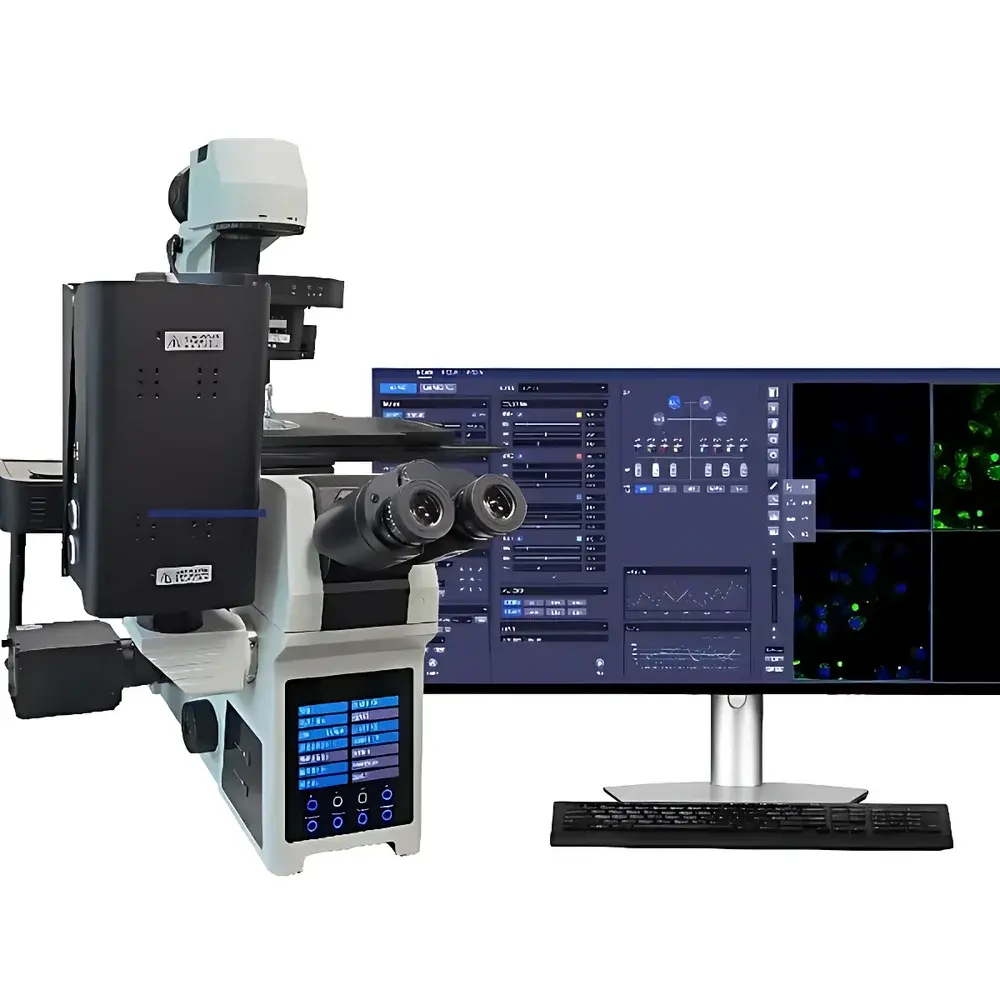
SHNTI HMS High-Spectral Microscopy System
| Brand | SHNTI |
|---|---|
| Origin | Shanghai, China |
| Model | HMS |
| Optical Resolution | 240 nm (XY), 600 nm (Z) |
| Camera Sensor | 2048 × 2048 back-illuminated sCMOS |
| Spatial Dimensions | XYZλt (5D acquisition) |
| Application Scope | Cellular biology, nanobiology, clinical pathology, materials science |
| Compliance Context | Designed for GLP-compliant research environments |
Overview
The SHNTI HMS High-Spectral Microscopy System is an advanced research-grade instrument engineered to unify spectral fingerprinting with diffraction-limited optical microscopy. Based on push-broom or tunable-filter-based hyperspectral imaging architecture—integrated into a motorized inverted or upright microscope platform—the system captures spatially registered reflectance, fluorescence, or transmission spectra across contiguous narrowband spectral channels (typically 400–1000 nm, configurable). Unlike conventional widefield or confocal microscopes that deliver monochromatic or RGB-limited contrast, the HMS acquires full spectral cubes (x, y, λ) at each voxel, enabling label-free biochemical mapping of endogenous chromophores (e.g., hemoglobin, melanin, NADH, lipids) and exogenous nanomaterials such as CeO₂ nanoparticles in live or fixed biological specimens. Its core measurement principle relies on wavelength-resolved photon detection coupled with precision piezo-driven Z-stacking, facilitating quantitative 3D spectral tomography without physical sectioning.
Key Features
- Nanometer-scale optical resolution: 240 nm lateral (XY) and 600 nm axial (Z) resolution, achieved via high-NA objective coupling, aberration-corrected spectral optics, and sub-pixel registration algorithms.
- Back-illuminated sCMOS detector: 2048 × 2048 pixel sensor with >80% quantum efficiency at 550 nm, low read noise (90 dB), optimized for low-light hyperspectral acquisition.
- Automated 5D data acquisition: Synchronized control of X-Y-Z stage, spectral filter tuning (motorized liquid crystal tunable filter or acousto-optic tunable filter), and time-lapse triggering enables XYZλt hyperspectral volumetric time-series capture.
- AI-powered virtual staining engine: Trained on histopathologically annotated spectral libraries, the embedded deep learning module (CNN-based spectral unmixing) generates synthetic H&E, trichrome, or immunofluorescence-like overlays directly from native spectral data—eliminating chemical fixation, sectioning artifacts, and photobleaching risks.
- Modular optical path design: Compatible with standard Nikon, Olympus, or Zeiss C-mount interfaces; supports transmitted light, epi-fluorescence, darkfield, and polarization contrast modalities.
Sample Compatibility & Compliance
The HMS accommodates diverse specimen formats including unstained tissue sections (4–20 µm), live-cell monolayers in glass-bottom dishes, suspended nanoparticles in aqueous media, and thin-film material samples on silicon wafers or ITO substrates. It operates within Class I laser safety limits when configured with LED or halogen broadband illumination; optional laser lines (e.g., 488 nm, 640 nm) comply with IEC 60825-1:2014. Data handling adheres to principles aligned with Good Laboratory Practice (GLP) frameworks: raw spectral cubes are saved in vendor-agnostic, metadata-rich formats (e.g., BSQ or BIL ENVI headers), supporting traceability. When deployed with SHNTI’s optional validated software suite, the system provides electronic signatures, user access logs, and immutable audit trails compliant with FDA 21 CFR Part 11 for regulated preclinical studies.
Software & Data Management
The HMS ships with SHNTI Hyperspectral Studio—a cross-platform application built on Qt and Python (NumPy, SciPy, scikit-learn). It delivers real-time spectral cube visualization, principal component analysis (PCA), vertex component analysis (VCA), and constrained energy minimization (CEM) for endmember extraction. Batch processing pipelines support radiometric calibration, flat-field correction, atmospheric compensation (for reflective mode), and spectral library matching against NIST-traceable reference databases. Export options include TIFF stacks, CSV spectral profiles, and MATLAB-compatible .mat files. API access (RESTful endpoints and Python SDK) enables integration into laboratory information management systems (LIMS) and automated analysis workflows.
Applications
- Cellular phenotyping: Discrimination of apoptosis, necrosis, and autophagy states via intrinsic fluorescence spectral shifts in NAD(P)H/FAD redox ratios.
- Nanobiology: Quantitative 3D tracking and spectral deconvolution of CeO₂ nanoparticle uptake kinetics and intracellular distribution in tumor cell lines (validated in MCF-7 and A549 models).
- Digital pathology: Label-free diagnosis of early-stage dysplasia in oral mucosa biopsies using spectral entropy and scattering slope metrics correlated with histological grade.
- Materials characterization: Mapping of grain boundaries, defect states, and dopant distributions in perovskite thin films through photoluminescence excitation (PLE) hyperspectral imaging.
- Pharmaceutical QA: Non-destructive assay of tablet coating uniformity and API crystallinity via near-infrared (NIR) reflectance hyperspectral imaging (900–1700 nm extension optional).
FAQ
What spectral range does the standard HMS configuration cover?
The base system operates from 400 nm to 1000 nm with 2–5 nm spectral sampling resolution, adjustable via hardware filter selection.
Is the system compatible with third-party microscope frames?
Yes—mechanical and optical interfaces follow ISO 8039 standards; adapter kits are available for Nikon Eclipse, Olympus IX, and Zeiss Axio series.
Can spectral data be exported for external machine learning training?
Absolutely: raw hypercubes are stored in open-format ENVI headers with embedded calibration metadata, fully interoperable with TensorFlow, PyTorch, and ENVI Classic.
Does the AI virtual staining module require cloud connectivity?
No—all inference runs locally on the host workstation; no patient data leaves the instrument network perimeter.
What maintenance protocols ensure long-term spectral calibration stability?
Built-in daily auto-calibration routines use onboard NIST-traceable tungsten-halogen and mercury-argon references; annual verification against PTB-certified standards is recommended.


