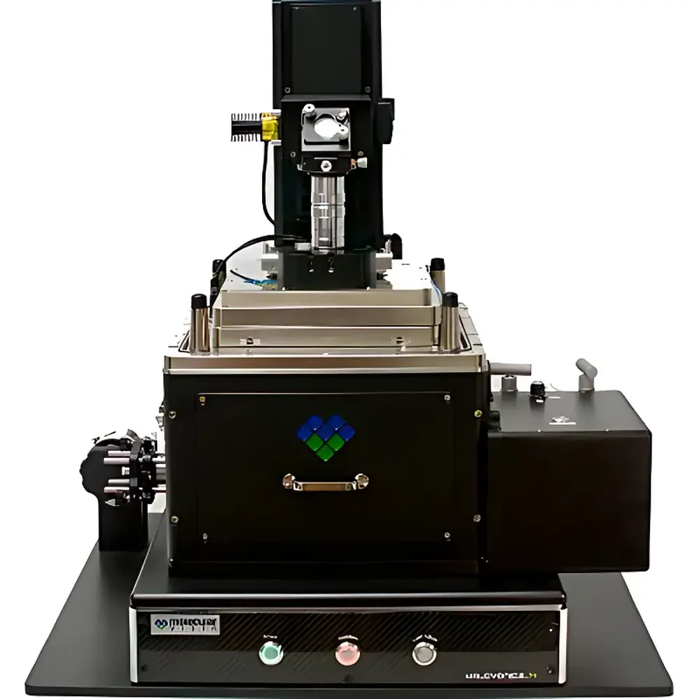
Molecular Vista VistaScope AFM-Visible-IR-Raman Correlative Microscopy System
| Brand | Molecular Vista |
|---|---|
| Origin | USA |
| Manufacturer Type | Authorized Distributor |
| Origin Category | Imported |
| Model | VistaScope |
| Price Range | USD 420,000 – 700,000 |
| Instrument Type | Material Science AFM |
| Position Detection Noise | ≤ 50 pm RMS |
| Sample Dimensions | Ø ≤ 25 mm, Thickness ≤ 10 mm |
| XY Stage Travel Range | 6 mm × 6 mm |
Overview
The Molecular Vista VistaScope is a correlative nanoscale imaging and spectroscopy platform integrating atomic force microscopy (AFM) with visible, infrared, and Raman modalities within a single, unified instrument architecture. At its core lies patented Photo-induced Force Microscopy (PiFM), a near-field technique that detects dipole–dipole interactions between a metallic AFM tip and optically excited sample regions—bypassing diffraction limits and thermal expansion artifacts inherent in conventional photothermal AFM methods. Unlike far-field optical detection or scanning near-field optical microscopy (s-SNOM), PiFM directly maps local electromagnetic field distributions without requiring external spectrometers or broadband detectors. This enables true sub-10 nm spatial resolution chemical imaging across the visible (400–800 nm), near-infrared (800–1200 nm), and mid-infrared (1000–4000 cm⁻¹) spectral ranges. The VistaScope’s co-aligned optical paths support top-, side-, and bottom-side illumination—critical for transparent, opaque, and multilayered samples—and enable seamless integration with external Raman spectrometers for tip-enhanced Raman spectroscopy (TERS) and surface-enhanced Raman scattering (SERS) characterization.
Key Features
- Sub-10 nm spatial resolution chemical imaging via PiFM across visible, NIR, and mid-IR spectral bands—no far-field optics or cryogenic cooling required.
- Integrated multi-modal acquisition: simultaneous or sequential AFM topography, PiFM amplitude/phase, NanoIR spectra, NanoVis absorption maps, and TERS mapping.
- Optomechanically stable, inverted–epi dual-path optical design enabling polarization-controlled excitation from three orthogonal directions (top, side, bottom).
- Low-noise position detection system with ≤ 50 pm RMS noise floor, optimized for high-fidelity force gradient sensing under modulated optical excitation.
- 6 mm × 6 mm closed-loop XY piezo stage with sub-nanometer step resolution and integrated Z feedback for dynamic height tracking during spectroscopic rastering.
- Modular architecture supporting third-party Raman spectrometers (e.g., Horiba, WITec, Renishaw) and optional s-SNOM phase detection add-on for complementary near-field optical phase contrast.
Sample Compatibility & Compliance
The VistaScope accommodates standard SEM/TEM-compatible substrates—including Si/SiO₂ wafers, CaF₂ IR windows, ITO-coated glass, and Au-coated mica—with maximum sample dimensions of Ø 25 mm and thickness ≤ 10 mm. Its open-access optical configuration permits in situ environmental control (e.g., gas cells, heating stages) and electrochemical cell integration. All PiFM and TERS data acquisition workflows comply with GLP/GMP documentation standards, including full audit trails, user authentication, and electronic signature support per FDA 21 CFR Part 11. Spectral calibration traceability follows NIST-traceable reference standards (e.g., polystyrene film for IR, silicon wafer for Raman), and measurement repeatability meets ASTM E2937-22 requirements for nanoscale chemical imaging validation.
Software & Data Management
VistaScope Control Suite v4.x provides unified control of AFM, PiFM, and Raman acquisition modules via a Python-extendable GUI. Real-time spectral stitching, hyperspectral unmixing (using non-negative matrix factorization), and pixel-wise fitting of IR absorption peaks (e.g., C=O stretch at 1720 cm⁻¹, Si–O–Si at 1080 cm⁻¹) are performed on-board using GPU-accelerated libraries. Raw datasets are stored in HDF5 format with embedded metadata (wavelength, laser power, modulation frequency, tip resonance shift), ensuring FAIR (Findable, Accessible, Interoperable, Reusable) compliance. Export options include standardized formats (JCAMP-DX for IR, SPE for Raman) compatible with commercial chemometric packages (e.g., GRAMS/AI, MATLAB Chemometrics Toolbox). Remote operation and multi-user scheduling are supported via secure TLS-encrypted web interface.
Applications
- Materials Science: Nanoscale mapping of polymer blend phase separation (e.g., PS/PMMA), zeolite framework vibrations (ZSM-5 Al/Si ratio gradients), and 2D material heterostructure strain fields (MoS₂, h-BN).
- Catalysis Research: In situ tracking of coke deposition dynamics during methanol-to-hydrocarbons (MTH) reactions via C=C stretch (1480 cm⁻¹) distribution mapping on catalyst surfaces.
- Nanophotonics: Quantitative LSPR field mapping of plasmonic nanostructures (Au–Al dimers, Ag nanoparticle arrays) with <3.1 nm resolution, validated against FDTD simulations.
- Life Sciences: Single-molecule absorption imaging of organic dyes (6-TAMRA) and protein–ligand binding interfaces using visible PiFM; correlative AFM–fluorescence–TERS of membrane protein clusters.
- Energy Materials: Chemical state evolution at solid–electrolyte interphases (SEI) in Li-ion batteries, cross-sectional analysis of perovskite solar cell layer composition, and defect-associated vibrational modes in quantum dot films.
FAQ
What distinguishes PiFM from conventional AFM-based infrared techniques like AFM-IR or photothermal FTIR?
PiFM measures optical force gradients arising from tip–sample dipole coupling, eliminating reliance on sample thermal expansion. This avoids lateral resolution degradation caused by heat diffusion—enabling consistent sub-10 nm resolution across all IR bands.
Can the VistaScope perform time-resolved pump–probe measurements?
Yes—the platform supports synchronized femtosecond laser systems (e.g., Ti:sapphire oscillators) for transient absorption imaging, with delay-stage integration and lock-in detection of photoinduced force transients.
Is TERS quantification possible with this system?
Quantitative TERS requires calibrated enhancement factors; VistaScope provides the necessary field-mapping capability (via PiFM) to locate hotspots and correlate local field intensity with measured Raman enhancement, fulfilling ISO/IEC 17025 metrological traceability requirements.
Does the system support vacuum or low-temperature operation?
The base configuration operates in ambient air or inert gas environments; optional UHV-compatible and cryogenic (4 K) stages are available through Molecular Vista’s engineering services group.
How is spectral calibration verified during routine use?
Automated daily calibration routines employ built-in reference targets (polystyrene, silicon, neon lamp) and cross-validate against NIST-traceable spectral databases embedded in the software—generating calibration reports compliant with ISO/IEC 17025 clause 5.9.


