JP-K3300 Chemiluminescence Gel Imaging System
| Brand | JiaPeng |
|---|---|
| Origin | Shanghai, China |
| Manufacturer Type | Authorized Distributor |
| Product Category | Domestic |
| Model | JP-K3300 |
| Instrument Type | Chemiluminescence Gel Imaging System |
| CCD Resolution | 9 MP (3000 × 3000) |
| Bit Depth | 16-bit |
| Dynamic Range | 4.8 OD (0–65,535 grayscale levels) |
| Pixel Binning Options | 1×1, 2×2, 3×3, 4×4, 6×6, 8×8 |
| Detection Sensitivity | ≥0.02 ng Protein |
| Signal-to-Noise Ratio | 56 dB |
| Lens Aperture | f/0.95 Fixed Focus Lens |
| CCD Sensor | Sony IMX533CLK-D, 4/3″ format |
| Cooling Temperature | −45 °C (ΔT ≈ −65 °C vs. ambient) |
| Quantum Efficiency | >75% |
| Pixel Size | 5.4 µm × 5.4 µm |
| White Light Transilluminator | Uniform top-mounted LED source |
| Dual Imaging Area | 9.5 × 9.5 cm and 12.5 × 12.5 cm |
| Power Supply | AC 220 V, 50/60 Hz |
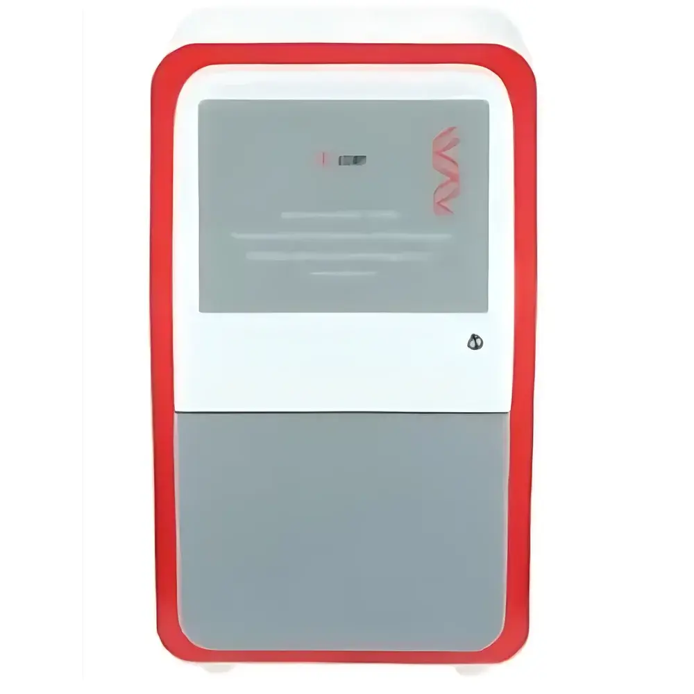
| Dimensions (W × D × H) | 390 × 440 × 735 mm |
| Weight | 18.3 kg |
Overview
The JP-K3300 Chemiluminescence Gel Imaging System is a fully integrated, automated platform engineered for high-sensitivity digital detection and quantitative analysis of nucleic acids and proteins in electrophoretic gels, blots, and luminescent assays. It operates on the principle of low-light photon capture using a thermoelectrically cooled scientific CMOS sensor, optimized for chemiluminescent (CL), bioluminescent (BL), and fluorescence-based detection modalities—including UV-transilluminated ethidium bromide-stained gels and visible-light-imaged membranes. The system’s optical architecture centers on a high-quantum-efficiency 4/3″ Sony IMX533CLK-D sensor with deep-cooling capability (−45 °C nominal, up to −65 °C ΔT), enabling ultra-low dark current (<0.0005 e⁻/pixel/s) and extended exposure times without thermal noise accumulation. Combined with an f/0.95 fixed-focus lens—among the widest apertures commercially available for benchtop imaging—the JP-K3300 delivers exceptional photon collection efficiency critical for detecting faint signals from low-abundance targets such as phosphorylated kinases or rare transcription factors.
Key Features
- Thermoelectrically cooled scientific-grade sensor (Sony IMX533CLK-D) with programmable temperature stabilization at −45 °C, ensuring reproducible signal acquisition across multi-hour exposures.
- f/0.95 ultra-fast fixed-focus lens optimized for maximum light throughput; optional f/0.8 lens upgrade available for enhanced sensitivity in ultra-low-light applications.
- Dual-stage imaging area (9.5 × 9.5 cm and 12.5 × 12.5 cm) accommodates standard mini- and midi-gels, Western blot membranes (including PVDF and nitrocellulose), and multi-well plates.
- Top-mounted uniform white LED transilluminator with calibrated intensity control, supporting both Coomassie- and silver-stained gel documentation under visible light.
- Motorized drawer-style dual-position sample stage with manual fine-focus adjustment; optional computer-controlled Z-axis lift platform available for automated focus calibration and batch processing.
- Programmable auto-shutdown for illumination sources (0–60 min timer), reducing photobleaching risk and extending LED service life.
- Anti-scatter sample tray design minimizes stray light interference, improving contrast and quantitative linearity in densitometric analysis.
Sample Compatibility & Compliance
The JP-K3300 supports a broad range of molecular biology workflows, including SDS-PAGE, agarose/TBE gels, Western blots (ECL, SuperSignal, Luminata), Northern/Southern blots, and luciferase reporter assays. Its 16-bit dynamic range (0–65,535) and 4.8 OD linear response enable accurate quantification across three orders of magnitude—meeting requirements for semi-quantitative and relative quantitation per ISO/IEC 17025 and CLSI EP17-A2 guidelines. The system’s firmware and acquisition software comply with ALCOA+ principles (Attributable, Legible, Contemporaneous, Original, Accurate) and support audit-trail-enabled operation suitable for GLP and GMP environments. While not FDA 21 CFR Part 11–certified out-of-the-box, the platform is configurable with electronic signature modules and user-access controls to meet regulatory documentation standards upon validation.
Software & Data Management
The JP-K3300 ships with proprietary imaging software compatible with Windows 10/11 (64-bit), featuring real-time preview, multi-channel overlay, background subtraction, lane/band detection, molecular weight calibration, and integrated densitometry. Raw image data are saved in lossless TIFF format with embedded metadata (exposure time, binning mode, lens ID, temperature log). Batch processing scripts allow standardized analysis across replicate experiments, while export options include CSV, Excel, and publication-ready PNG/PDF. The software supports DICOM export for institutional PACS integration and includes built-in tools for MIQE-compliant qPCR gel documentation and REACH-relevant protein expression reporting.
Applications
- Quantitative Western blotting with ECL substrates (e.g., HRP-conjugated secondary antibodies)
- Detection of low-copy-number DNA fragments post-PCR or restriction digest
- Analysis of post-translational modifications (e.g., phospho-specific antibodies) requiring high signal-to-noise ratios
- Luciferase-based promoter activity assays and cell viability screening
- Documentation and comparative analysis of Coomassie- and silver-stained 2D gels
- Verification of CRISPR/Cas9 editing efficiency via T7E1 or Surveyor nuclease assays
- Quality control of recombinant protein purification batches using SDS-PAGE densitometry
FAQ
What is the minimum detectable amount of protein using the JP-K3300?
The system achieves ≥0.02 ng sensitivity for standard chemiluminescent detection of horseradish peroxidase (HRP)-labeled proteins under optimized conditions (e.g., enhanced ECL substrate, 5-min exposure, 4×4 binning).
Can the JP-K3300 perform fluorescence imaging?
No—it lacks excitation light sources and emission filters required for fluorescence; it is dedicated to chemiluminescence, bioluminescence, and visible-light transillumination.
Is the software compliant with 21 CFR Part 11?
The base software does not include electronic signatures or audit trails by default, but validated add-on modules are available for regulated environments upon request and site-specific qualification.
What cooling method is used for the sensor?
A multi-stage Peltier cooler maintains stable −45 °C sensor temperature, with active heat dissipation via aluminum heatsink and low-noise fans.
Does the system support time-lapse luminescence imaging?
Yes—programmable exposure sequencing (up to 99 frames) enables kinetic monitoring of luciferase activity or decay kinetics of unstable CL substrates.
Related Products


