
RWD MOIS HT Small Animal In Vivo Optical Imaging System
| Brand | RWD |
|---|---|
| Origin | Guangdong, China |
| Manufacturer Type | Manufacturer |
| Origin Category | Domestic |
| Model | MOIS HT |
| Instrument Type | Optical Imaging System |
| Animal Type | Small Rodents (e.g., mice, rats) |
| Scan Resolution | 1024 × 1024 pixels |
| Field of View (FOV) | Up to 25 cm × 25 cm (optional) |
| Excitation/Emmission Filter Set | 19-position automated filter wheel (7 emission + 12 excitation filters) |
Overview
The RWD MOIS HT Small Animal In Vivo Optical Imaging System is a high-sensitivity, multi-modal preclinical optical imaging platform engineered for quantitative longitudinal studies in live rodents. It operates on the principles of bioluminescence detection (photon counting under ultra-low-light conditions), fluorescence reflectance/imaging (excitation-emission spectral separation), Cherenkov radiation imaging (for radionuclide tracking), and integrated X-ray anatomical referencing. Designed for rigorous biomedical research environments—including oncology, immunology, infectious disease, and drug development—the system delivers reproducible, calibration-traceable photon flux measurements with sub-picomolar sensitivity. Its back-illuminated scientific-grade CCD detector, cooled to −90 °C, ensures minimal dark current noise and quantum efficiency exceeding 90% across the 500–700 nm spectral window—critical for resolving weak, spatially localized signals from deep-tissue reporters or early-stage lesions.
Key Features
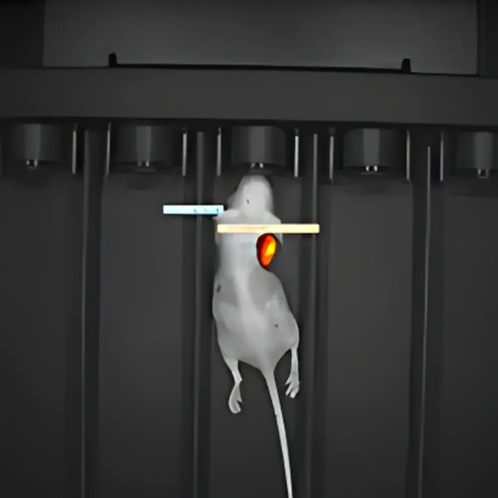
- Multi-Modal Acquisition Architecture: Seamless integration of bioluminescence imaging (BLI), fluorescence imaging (FLI), Cherenkov luminescence imaging (CLI), and co-registered X-ray radiography—enabling cross-modal validation and anatomical context mapping without subject repositioning.
- Ultra-Low-Noise Detection: Scientific CCD sensor with thermoelectric cooling to −90 °C; F/0.95 lens assembly optimized for maximum photon throughput; 1024 × 1024 pixel resolution with pixel binning support for enhanced signal-to-noise ratio (SNR) in low-flux applications.
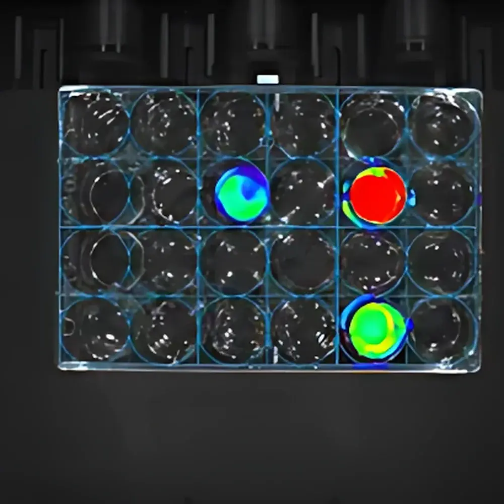
- Spectral Unmixing Capability: Software-enabled linear unmixing using user-defined reference spectra to resolve overlapping fluorophore emissions (e.g., GFP vs. Cy5.5) and suppress auto-fluorescence contributions—essential for multiplexed probe studies in murine models.
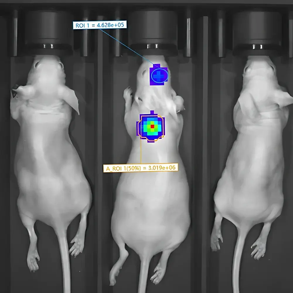
- Automated Calibration & Drift Compensation: Onboard dark-current and readout-bias correction routines executed prior to each acquisition session; standardized radiometric units (photons/sec/cm²/sr) applied across all imaging modes to ensure inter-session and inter-laboratory data comparability.
- High-Throughput Workflow Support: 5-channel animal chamber with independent temperature control (20–40 °C, ±0.1 °C stability) and calibrated isoflurane anesthesia delivery (0.5–5.0% adjustable); synchronized multi-animal sequential imaging reduces per-subject handling time and physiological variability.
Sample Compatibility & Compliance
The MOIS HT accommodates standard laboratory rodents (mice and rats, up to 50 g) in supine or prone positioning. All optical modules comply with IEC 61000-4 electromagnetic compatibility standards. The system supports GLP-compliant workflows through audit-trail-enabled software logging (user actions, acquisition parameters, calibration timestamps). While not FDA-cleared for diagnostic use, it meets ASTM E2594-18 guidelines for quantitative optical imaging instrumentation validation and is routinely deployed in academic and pharmaceutical research settings adhering to ISO/IEC 17025 quality management requirements.
Software & Data Management
Acquisition and analysis are managed via RWD’s proprietary MOIS Studio software suite—comprising real-time online acquisition console and offline quantitative workstation modules. The acquisition interface supports time-series imaging, multi-mode sequential triggering (e.g., BLI → FLI → X-ray), and hardware-synchronized event logging. Offline analysis includes region-of-interest (ROI) quantification, spectral unmixing, kinetic curve fitting, and export of MIAME-compliant metadata. Optional molecular quantification module enables absolute flux calibration using NIST-traceable light sources and certified fluorescent standards. Data export formats include TIFF (16-bit), CSV, and DICOM-SR for PACS integration.
Applications
- Tumor xenograft growth and metastasis monitoring via luciferase-expressing cell lines
- In vivo evaluation of targeted fluorescent probes (e.g., protease-activated agents, pH-sensitive dyes)
- Immunotherapy response assessment using dual-reporter T-cell trafficking models
- Longitudinal tracking of radiolabeled therapeutics via Cherenkov signal detection
- Structural-functional correlation through fused X-ray/optical overlays for precise anatomical registration
- Pharmacokinetic and biodistribution studies requiring high temporal resolution and inter-subject normalization
FAQ
What is the minimum detectable photon flux for bioluminescence imaging?
The system achieves sub-100 photons/sec/cm²/sr detection limits under optimal conditions (−90 °C sensor, 5-minute integration, F/0.95 lens, no binning). Actual sensitivity depends on reporter expression level, tissue depth, and background autofluorescence.
Does the system support spectral unmixing for more than two fluorophores simultaneously?
Yes—up to four spectrally distinct fluorophores can be resolved using reference spectra imported from vendor-provided or empirically acquired emission profiles.
Is the X-ray module integrated or add-on?
The X-ray imaging capability is fully integrated into the MOIS HTX variant; the base MOIS HT model does not include X-ray hardware but retains mechanical and software interfaces for future field upgrade.
How is temperature stability maintained during long-duration acquisitions?
Each animal channel features independent Peltier-based thermal regulation with closed-loop feedback control, maintaining setpoint accuracy within ±0.1 °C over sessions exceeding 60 minutes.
Can raw image data be exported for third-party analysis (e.g., MATLAB, Python)?
Yes—TIFF stacks with embedded EXIF metadata (exposure time, binning factor, filter position, gain setting) are natively supported; calibration coefficients and spectral response curves are provided in JSON format for custom processing pipelines.


