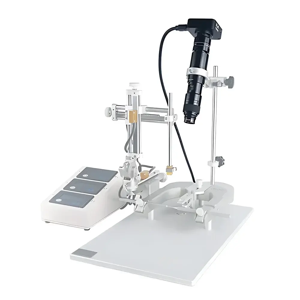
RWD 77001D Digital Surgical Microscope for Stereotactic Brain Surgery
| Brand | RWD |
|---|---|
| Origin | Shenzhen, China |
| Manufacturer Type | OEM/ODM Manufacturer |
| Country of Origin | China |
| Model | 77001D |
| Pricing | Upon Request |
| Sensor | 1/2.33" Panasonic 16 MP CMOS |
| Pixel Size | 1.335 µm × 1.335 µm |
| HDMI Output | 1920×1080 @ 60 fps |
| Lens Mount | C/CS |
| Storage | microSD up to 64 GB |
| Input Voltage | DC 5–12 V |
| Focus Travel | Manual 8 mm |
| Magnification Range | 20×–120× (Optical Zoom Ratio: 0.18–1.13×) |
| Working Distance | 180 mm |
| Depth of Field | 0.41 mm (high mag) to 6.7 mm (low mag) |
| Resolution | 7.6 µm (high mag), 19.8 µm (low mag) |
| FOV (diagonal, 1/3″ sensor) | 5.3 mm (high mag) to 33.3 mm (low mag) |
| On-Screen Tools | 8 adjustable annotation lines + center crosshair |
| Exposure Control | Manual/Auto/One-Touch with compensation |
| White Balance | Manual/Auto/One-Touch with RGB channel adjustment |
| UI Language | English & Chinese |
| Camera Interface | C-mount |
Overview
The RWD 77001D Digital Surgical Microscope is an integrated optical-imaging platform engineered specifically for stereotactic neurosurgery, intracranial injection procedures, and small-animal surgical interventions. Unlike conventional binocular microscopes requiring prolonged head-down posture and ocular strain, this system employs a high-resolution CMOS imaging core coupled with variable-magnification optics to project real-time, high-fidelity video directly onto an external display. Its design adheres to the ergonomic and operational requirements of preclinical neuroscience labs—enabling hands-free, collaborative visualization while maintaining precise spatial orientation relative to bregma/lambdа coordinates. The system operates on the principle of coaxial brightfield imaging with optimized depth-of-field management across its 20×–120× magnification range, delivering consistent resolution and geometric fidelity without mechanical parallax. It does not replace intraoperative navigation systems but serves as a critical visual verification layer during craniotomy, dural incision, electrode placement, or viral vector injection—ensuring anatomical accuracy prior to irreversible tissue manipulation.
Key Features
- Full-digital imaging workflow: Eliminates eyepiece dependency; supports simultaneous multi-user observation via HDMI 1080p60 output to clinical monitors or teaching displays.
- Variable optical magnification (20×–120×) with calibrated zoom ring and fixed 180 mm working distance—optimized for compatibility with standard stereotaxic frames (e.g., David Kopf, Stoelting).
- High-depth-of-field optics: 0.41 mm at 120× to 6.7 mm at 20×, enabling clear focus across layered cortical structures without constant refocusing during needle advancement.
- Ergonomic articulation: Adjustable microscope body allows vertical, oblique, and lateral repositioning—critical for accessing posterior fossa or lateral ventricles in rodent models.
- On-device image annotation: Eight user-configurable measurement lines (color, thickness, position) plus toggleable center crosshair for coordinate referencing and trajectory alignment.
- Dual exposure control architecture: Combines auto-exposure with manual override and one-touch exposure lock—essential for managing dynamic contrast shifts during blood vessel visualization or fluorescent tracer injection.
- Robust data capture: Internal microSD slot (up to 64 GB) supports timestamped still capture and lossless H.264 video recording—fully traceable for GLP-compliant experimental documentation.
Sample Compatibility & Compliance
The RWD 77001D is validated for use with murine (C57BL/6, BALB/c), rat (Sprague-Dawley, Wistar), and ferret models undergoing stereotactic surgery. Its 180 mm working distance accommodates standard skull-mounted headplates and permits unobstructed access for micromanipulator-controlled injection needles (e.g., Nanoject III, Drummond). All optical components comply with ISO 10993-5 biocompatibility standards for non-invasive device surfaces. While not FDA-cleared as a medical device, the system meets ISO 13485-aligned manufacturing controls per RWD’s QMS certification. Data export formats (JPEG, MP4) are compatible with institutional PACS archives and support audit-ready metadata embedding (date/time, magnification, exposure settings) required under GLP Annex 11 and 21 CFR Part 11–compliant environments when paired with validated storage protocols.
Software & Data Management
No proprietary desktop software is required for basic operation—the device functions as a UVC-compliant USB video class (UVC) peripheral when connected to Windows/Linux hosts. For advanced functionality, RWD provides optional firmware-upgradable firmware modules supporting DICOM-conformant image export, frame-averaged low-light enhancement, and ROI-based histogram analysis. All captured media include embedded EXIF metadata (magnification factor, exposure time, white balance mode, lens ID). Remote control via IR handset enables hands-free shutter activation and menu navigation—reducing contamination risk during sterile procedures. Firmware updates are delivered via signed .bin files with SHA-256 checksum validation to ensure integrity and version traceability.
Applications
- Stereotactic intracerebral injection of AAV, lentivirus, or cell suspensions into hippocampus, striatum, or prefrontal cortex.
- Real-time guidance during cranial window implantation and two-photon surgery preparation.
- Post-operative wound assessment and suture line evaluation in longitudinal behavioral studies.
- Training module for graduate students: Enables instructor-led demonstration of craniotomy technique with synchronized audio commentary and annotated playback.
- Quality assurance in contract research organizations (CROs): Standardized imaging protocol templates ensure inter-operator consistency across multi-site pharmacology studies.
FAQ
Is the RWD 77001D compatible with third-party stereotaxic frames?
Yes—it maintains full mechanical interoperability with all major brands (Kopf, Stoelting, Angle Two) via standard mounting adapters. The fixed 180 mm working distance ensures repeatable alignment across setups.
Can exposure settings be locked during time-lapse injection sequences?
Yes. One-touch exposure lock preserves gain and shutter parameters across sequential frames, preventing automatic brightness fluctuations during slow-infusion protocols.
Does the system support live measurement tools (e.g., distance, area) on screen?
No built-in calibratable measurement suite is included; however, the eight programmable annotation lines enable manual scale referencing when used with stage micrometers or graticule slides.
What is the maximum recommended cable length for stable HDMI 1080p60 transmission?
For guaranteed signal integrity, use certified High-Speed HDMI cables ≤ 5 meters. Active fiber-optic extenders are supported for distances up to 100 meters.
Is firmware update capability available over network or only via microSD?
Firmware updates are performed exclusively via microSD card—ensuring air-gapped security in regulated lab environments where network connectivity is restricted.


