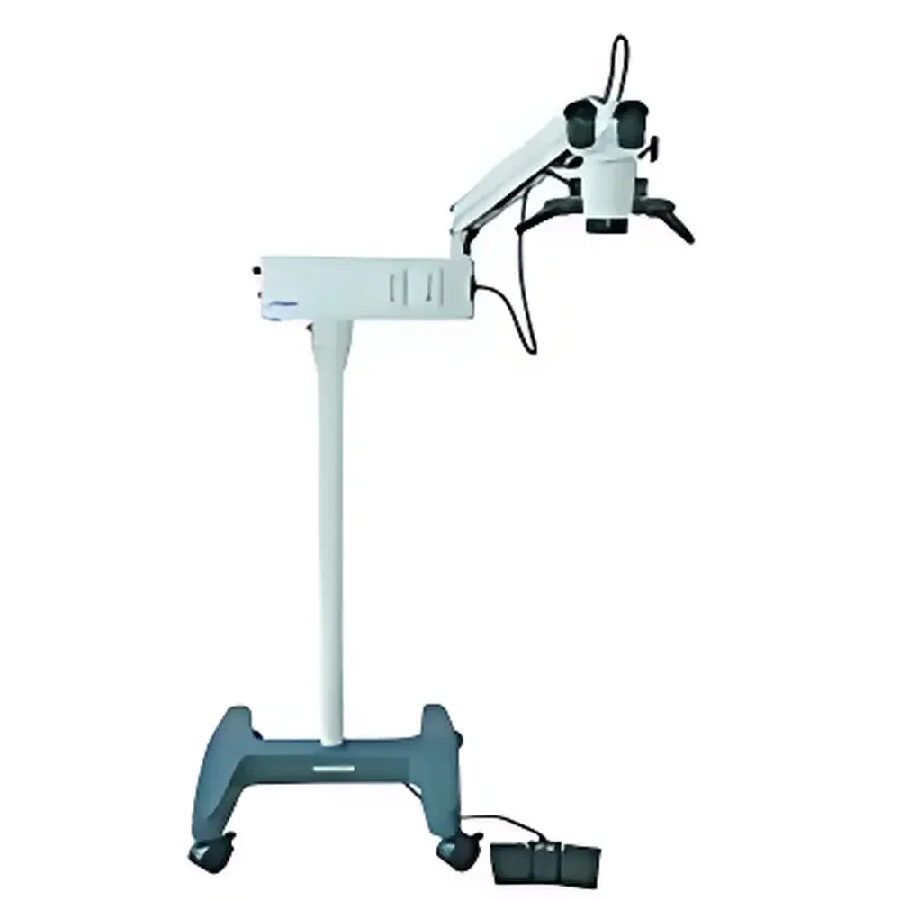
RWD 77002 Vertical Animal Surgical Microscope
| Brand | RWD |
|---|---|
| Origin | Guangdong, China |
| Manufacturer Type | OEM/ODM Manufacturer |
| Country of Origin | China |
| Model | 77002 |
| Pricing | Upon Request |
| Eyepiece Magnification | 12.5× |
| Objective Focal Length | 200 mm |
| Working Distance | 190 mm |
| Primary Magnification Steps | 5.3×, 8×, 12× |
| Field Diameter | 38 mm, 25 mm, 17 mm |
| Diopter Adjustment Range | ±5 D |
| Interpupillary Distance Adjustment | 50–70 mm |
| Illumination Source | 12 V / 100 W Cold-Reflective Medical Halogen Lamp |
| Illumination Geometry | 6° + 0° Coaxial Cold Light |
| Illuminance at Object Plane | ≥20,000 lx |
| Horizontal Arm Reach Radius | 870 mm |
| Fine Focus Travel | 30 mm |
| Input Voltage | AC 220 V ±10 %, 50 Hz ±1 Hz |
| Power Consumption | 120 VA |
| Fuse Rating | AC 250 V, T1.25 A |
| Net Weight | 41 kg |
Overview
The RWD 77002 Vertical Animal Surgical Microscope is a compact, dual-eye coaxial illumination surgical microscope engineered for precision in preclinical rodent and small-animal neurosurgery, ophthalmic procedures, vascular anastomosis, and microdissection. Designed around a robust vertical column architecture with a counterbalanced horizontal articulating arm, it delivers stable optical alignment and ergonomic positioning without floor footprint compromise. Its optical path employs a high-transmission achromatic objective lens system (200 mm focal length) coupled with fixed 12.5× wide-field eyepieces and a three-step zoom objective turret (5.3×, 8×, 12×), enabling rapid magnification switching while maintaining parfocality and minimal chromatic aberration across the entire field. The coaxial illumination system integrates a cold-reflective 12 V / 100 W halogen lamp (HLX64627) with dual-angle beam delivery (6° oblique + 0° axial), ensuring uniform, shadow-free illumination at ≥20,000 lx on the specimen plane—critical for minimizing thermal load on delicate neural or ocular tissues during prolonged exposure.
Key Features
- Vertical column design with 870 mm horizontal arm reach radius, supporting flexible positioning over stereotaxic frames, heating pads, or anesthesia systems without obstructing operator access.
- Three-step manual zoom objective (5.3×, 8×, 12×) with fixed 12.5× wide-field eyepieces, delivering effective magnifications of 66×, 100×, and 150×—optimized for both gross tissue orientation and fine suture visualization.
- Cold-light coaxial illumination system with dual-angle (6° + 0°) beam geometry, eliminating specular glare and preserving tissue viability during extended procedures.
- Precision fine-focus mechanism with 30 mm travel range and calibrated micrometer scale, enabling sub-millimeter depth adjustment under live observation.
- Ergonomic interpupillary adjustment (50–70 mm) and ±5 D diopter compensation per eyepiece, accommodating diverse user anthropometry and refractive requirements.
- Stable 41 kg base-weighted construction with vibration-dampening feet, ensuring optical stability even on shared lab benches adjacent to centrifuges or shakers.
Sample Compatibility & Compliance
The RWD 77002 is validated for use with murine, rat, rabbit, and avian models in both acute and chronic surgical settings. Its 190 mm working distance accommodates standard stereotaxic holders (e.g., David Kopf, Stoelting), head-fixation clamps, and intraoperative imaging accessories. All optical components comply with ISO 10940:2018 (Surgical Microscopes — Requirements and Test Methods) for resolution, illuminance uniformity, and thermal safety. The halogen lamp assembly meets IEC 60601-2-41:2015 for medical electrical equipment safety, including leakage current limits and mechanical stability under tilt testing. While not FDA-cleared as a Class II device (intended for human use), its design aligns with GLP-compliant preclinical facility standards—including traceable calibration logs, service history documentation, and compatibility with audit-ready maintenance records.
Software & Data Management
As a hardware-centric surgical platform, the 77002 operates without embedded firmware or proprietary software. However, it supports seamless integration into regulated digital workflows via optional C-mount interfaces (RWD 77017, 1/2″ standard) compatible with FDA-listed scientific cameras (e.g., Canon EOS DSLRs with tethering via EOS Utility or third-party SDKs). When paired with compliant imaging software (e.g., NIS-Elements, MetaMorph, or ImageJ/Fiji with audit-trail plugins), the system enables timestamped image capture, metadata embedding (magnification, illumination intensity, date/time), and export in DICOM-conformant or TIFF formats suitable for GLP/GCP study archives. Optional trinocular head (RWD 77029) facilitates simultaneous operator viewing and real-time teaching or remote supervision—fully compatible with HIPAA/FDA 21 CFR Part 11–compliant video management platforms when deployed with appropriate encryption and access controls.
Applications
- Microvascular surgery: End-to-end anastomosis of femoral or carotid arteries in rat models, requiring consistent 100×–150× magnification and stable illumination for suture placement.
- Neurosurgical interventions: Craniotomy-assisted cortical electrode implantation, optogenetic fiber insertion, or lesion induction with precise depth control enabled by the 30 mm fine-focus range.
- Ophthalmic procedures: Corneal suturing, lens extraction, or intravitreal injection in mice and rabbits, where cold illumination prevents corneal desiccation and thermal distortion.
- Transplantation models: Pancreatic islet or ovarian tissue grafting under visual guidance, leveraging the large field diameter (up to 38 mm at 5.3×) for donor tissue orientation.
- Training and education: Paired with trinocular configuration and HDMI capture, supports competency-based assessment in veterinary and biomedical graduate curricula aligned with AALAS training standards.
FAQ
Is the RWD 77002 certified for human clinical use?
No. It is designed and validated exclusively for preclinical animal research applications and does not hold FDA 510(k), CE Mark Class IIa, or MDR certification for human surgical use.
Can the microscope be integrated with electrophysiology rigs or behavioral tracking systems?
Yes. Its rigid column and horizontal arm allow unobstructed mounting above stereotaxic frames, patch-clamp setups, or operant chambers. Cable management grommets and accessory mounting threads (M4/M6) support synchronized hardware integration.
What is the recommended maintenance schedule for the cold-light source?
The HLX64627 halogen lamp should be replaced after 100 hours of cumulative operation or upon visible filament degradation; annual recalibration of illuminance output using a NIST-traceable photometer is advised for GLP compliance.
Does RWD provide installation qualification (IQ) or operational qualification (OQ) documentation?
Yes. Upon request, RWD supplies IQ/OQ templates aligned with ISO/IEC 17025 and ASTM E2500-13, including mechanical stability tests, illuminance mapping reports, and focus repeatability verification protocols.
Are replacement objectives available for higher magnification (e.g., 200× or 300×)?
Yes. RWD offers dedicated auxiliary objectives (77013: 200×, 77014: 250×, 77027: 300×, 77015: 400×) that maintain the same 190 mm working distance and are mechanically matched to the 77002’s zoom turret interface.
Related Products


