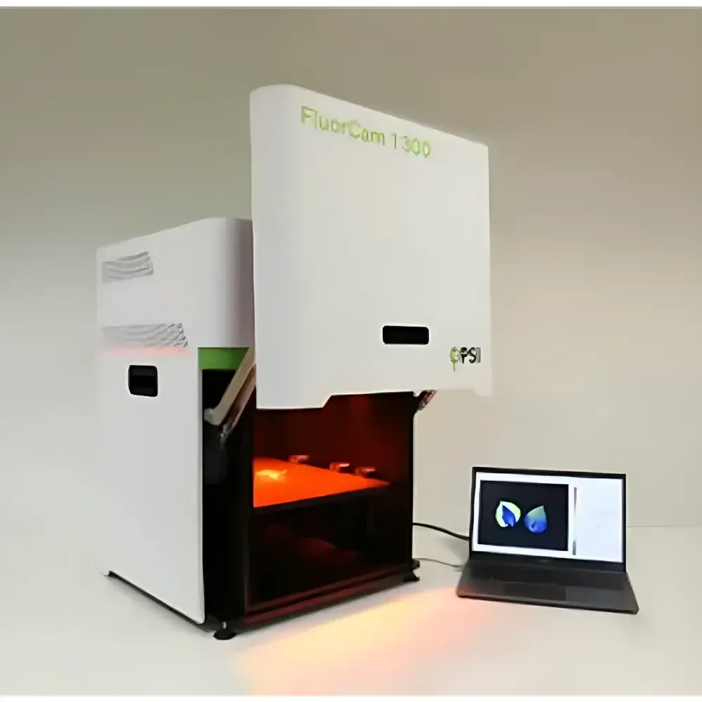
FluorCam Plant Photosynthetic Phenotyping Imaging System by PSI
| Brand | PSI (Czech Republic) |
|---|---|
| Origin | Czech Republic |
| Model Variants | Desktop, Floor-standing, Large Floor-standing |
| Excitation Light Sources | UV–NIR LED Array (365 nm, 450 nm, 470 nm, 490 nm, 520 nm, 590 nm, 630 nm, 735 nm, White Cold LED) |
| Fluorescence Detection Channels | Adjustable via Motorized Filter Wheel (FRET-compatible filters for GFP, RFP, YFP, BFP, CFP, SYBR Green, DAPI, etc.) |
| Imaging Area | Up to 35 × 35 cm |
| Sample Height Range | 20–100 cm (motorized adjustable stage) |
| Measurement Modes | PAM-modulated chlorophyll fluorescence imaging, OJIP transient kinetics imaging, multicolor steady-state fluorescence imaging, chlorophyll distribution mapping, fluorescent protein expression profiling, secondary metabolite fluorescence profiling |
| Software | FluorCam 7.0 with automated protocol scheduling, time-stamped data archiving, GLP-compliant audit trail, and FDA 21 CFR Part 11-ready user access control |
Overview
The FluorCam Plant Photosynthetic Phenotyping Imaging System is a high-resolution, multi-modal fluorescence imaging platform engineered by Photon Systems Instruments (PSI), Czech Republic, for non-invasive, quantitative analysis of photosynthetic performance and molecular phenotypes in living biological specimens. At its core, the system integrates pulse-amplitude modulated (PAM) chlorophyll fluorescence imaging with multicolor excitation–emission fluorescence spectroscopy, enabling simultaneous acquisition of physiological, biochemical, and genetic readouts across spatially resolved plant tissues and whole organisms. Its measurement principle relies on controlled photoexcitation of endogenous fluorophores—including chlorophyll a, NAD(P)H, phenolic compounds, and coumarins—as well as exogenous reporters such as GFP, RFP, and SYBR Green—followed by high-sensitivity, spectrally resolved detection using cooled CCD or sCMOS sensors. This dual-capability architecture supports both dynamic photophysiological assays (e.g., quantum yield of PSII, NPQ relaxation kinetics, OJIP transients) and static molecular phenotyping (e.g., promoter activity mapping, stress-induced metabolite accumulation, transgene expression gradients), making it suitable for functional genomics, abiotic/biotic stress physiology, breeding program screening, and algal biotechnology research.
Key Features
- Integrated multi-wavelength LED excitation array covering UV (365 nm) through far-red (735 nm), plus cold white LED simulating natural irradiance spectra—enabling selective activation of diverse fluorophores without thermal artifacts.
- Motorized filter wheel with ≥8 positions, pre-configured for standard fluorescent proteins (GFP, RFP, YFP) and nucleic acid dyes (DAPI, SYBR Green), with optional custom filter sets for BFP, CFP, or specialized probes.
- Three scalable configurations: Desktop (20 × 20 cm imaging area), Floor-standing (25 × 25 cm), and Large Floor-standing (35 × 35 cm), each featuring a motorized Z-axis sample stage (20–100 cm travel) for precise focal plane alignment across heterogeneous samples.
- Full software-driven hardware orchestration: All light sources, filters, shutter timing, and camera gain are synchronized and controlled via FluorCam 7.0—no manual reconfiguration required between measurement modes.
- Automated long-term phenotyping workflows: Users define protocols with programmable illumination regimes (intensity, duration, spectral composition), imaging intervals (seconds to days), and repetition cycles; all raw and processed data are timestamped and archived with metadata tagging.
- Robust optical design with uniform illumination homogeneity (>95% across FOV) and calibrated radiometric output—ensuring inter-experimental reproducibility essential for longitudinal studies and multi-lab collaborations.
Sample Compatibility & Compliance
The FluorCam system accommodates a broad spectrum of biological samples without fixation or sectioning: detached leaves, fruits, flowers, mosses, lichens, intact seedlings (Arabidopsis, tobacco), mature crop plants (wheat, maize), microalgae cultures (in droplets, multiwell plates, agar slants), macroalgae thalli, and selected animal models (e.g., Aplysia californica, Caenorhabditis elegans). Sample mounting is facilitated by standardized trays, petri dish holders, and adjustable clamps compatible with growth chambers and climate-controlled rooms. From a regulatory standpoint, FluorCam 7.0 supports Good Laboratory Practice (GLP) and Good Manufacturing Practice (GMP) workflows through role-based user authentication, electronic signatures, full audit trails, and compliance-ready documentation export—fully aligned with FDA 21 CFR Part 11 requirements for data integrity in regulated environments.
Software & Data Management
FluorCam 7.0 is a modular, Windows-based application designed for scientific rigor and operational efficiency. It provides real-time visualization of fluorescence kinetics, pixel-wise parameter mapping (e.g., Fv/Fm, ΦPSII, NPQ, qP), and batch processing of time-series datasets. The software includes built-in statistical tools for ROI-based quantification, normalization against reference standards, and export to MATLAB, Python (via HDF5), or spreadsheet formats. Raw image stacks are stored in lossless TIFF or PSI-native .FCM format, preserving full bit-depth and metadata (exposure time, gain, excitation wavelength, filter ID, timestamp). All user actions—including protocol edits, parameter adjustments, and report generation—are logged with ISO-compliant timestamps and operator IDs, ensuring traceability from acquisition to publication.
Applications
- High-throughput screening of mutant libraries for photosynthetic efficiency or stress tolerance traits.
- Time-resolved analysis of drought, salinity, heavy metal (e.g., Cd, Pb), or pathogen-induced photoinhibition dynamics.
- Functional validation of transcription factors (e.g., WRKY family) in stress-responsive gene networks using promoter-GFP fusions.
- Quantitative assessment of nanoparticle-mediated enhancement of disease resistance mechanisms in crops.
- In vivo monitoring of microalgal lipid accumulation and redox status under nutrient limitation or light stress.
- Non-destructive evaluation of fruit ripening, post-harvest senescence, and quality deterioration markers via autofluorescence shifts.
FAQ
What types of fluorescence measurements can be performed simultaneously?
The system supports concurrent acquisition of PAM chlorophyll fluorescence parameters (F0, Fm, Fv/Fm, ΦPSII) and steady-state multicolor fluorescence intensities (e.g., green/red/far-red ratios) under identical illumination conditions—enabling direct correlation between photosynthetic function and secondary metabolite distribution.
Is the system compatible with controlled-environment growth chambers?
Yes—standard configurations include chamber-integrated mounting brackets and cable routing ports; optional environmental sensor inputs (temperature, humidity, CO₂) allow synchronized logging with fluorescence data.
Can FluorCam data be integrated into LIMS or ELN platforms?
FluorCam 7.0 supports automated data export via scheduled tasks to network drives or REST API endpoints; structured metadata adheres to MIAPPE (Minimum Information About a Plant Phenotyping Experiment) standards for interoperability.
Does the system require routine calibration?
PSI provides factory-calibrated LED irradiance profiles and sensor quantum efficiency curves; users may perform optional daily dark-current and flat-field corrections via integrated routines to maintain long-term signal stability.
Are training and application support services available internationally?
PSI offers remote and on-site installation, method development workshops, and dedicated application scientist consultation—available globally through authorized distributors and direct technical support channels.


