WINDIAS Image Analysis System for Leaf Area and Plant Morphometrics
| Origin | UK |
|---|---|
| Manufacturer Type | Authorized Distributor |
| Origin Category | Imported |
| Model | WINDIAS Image Analysis System |
| Pricing | Available Upon Request |
| Maximum Scanning Area (Flatbed) | 400 × 370 mm |
| Maximum Scanning Area (Conveyor) | 250 × 290 mm |
| CCD Camera Resolution | 681 × 582 pixels |
| Area Measurement Accuracy | ≤4% |
| Frame Capture Rate | 1/25 s (50 Hz, real-time) |
| Software Image Resolution | 512 × 512 pixels, 24-bit color |
| Conveyor Speed Options | 60 / 100 / 140 / 190 mm/s |
| Max Conveyor Sample Width | 250 mm |
| Max Conveyor Sample Thickness | 5 mm |
| Camera Mount Height | 1150 mm |
| Flatbed Flattening Aid | Polypropylene sheet for curled leaf stabilization |
Overview
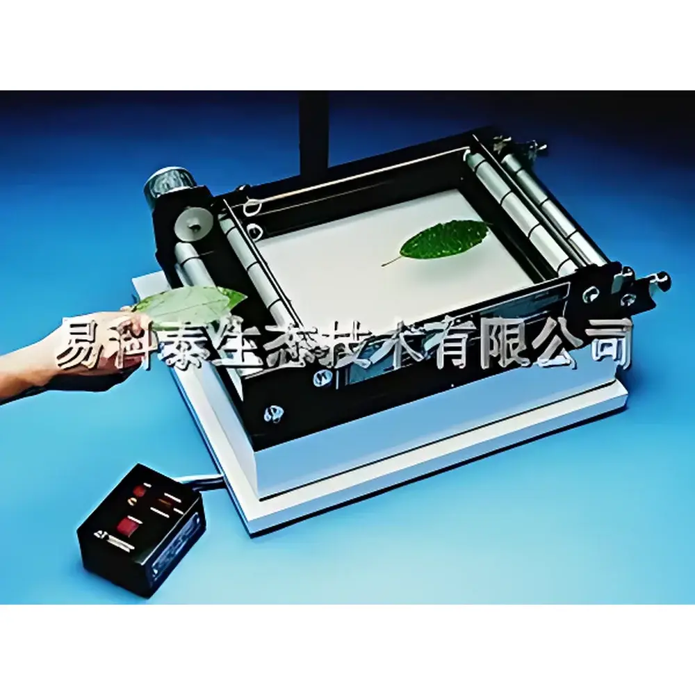
The WINDIAS Image Analysis System is a dedicated digital morphometric platform engineered for quantitative plant phenotyping in controlled laboratory and greenhouse environments. Designed specifically for researchers in plant physiology, agronomy, and ecological botany, the system employs high-fidelity digital image acquisition and pixel-based segmentation algorithms to deliver reproducible measurements of leaf area, lesion area, seed count, root tip density, and other two-dimensional structural parameters. Its core methodology relies on calibrated grayscale or RGB imaging under uniform illumination, followed by threshold-based binary conversion and region-of-interest (ROI) quantification—principles aligned with ISO 11540:2020 (Plant Phenotyping — Image-Based Measurement Protocols) and widely adopted in GLP-compliant crop screening workflows.
Key Features
- Dual-mode imaging architecture: supports both flatbed scanning (for pressed or flattened samples) and conveyor-based high-throughput acquisition (for unattended batch processing of detached leaves, seeds, or root segments)
- Integrated illumination system: top-mounted adjustable LED light source with diffused acrylic cover ensures uniform spectral distribution and minimizes specular reflection artifacts
- High-stability optical train: fixed-focus color CCD camera (681 × 582 effective pixels) mounted on a rigid 1150 mm-height vertical stand, optimized for consistent working distance and geometric fidelity
- Polypropylene flattening sheet included: mitigates parallax and curvature-induced measurement error in fresh, hydric, or senescing leaf specimens
- Real-time frame capture: WDIGC PCI image acquisition card enables synchronized 50 Hz (1/25 s) sampling, essential for motion artifact suppression during conveyor operation
- Configurable conveyor module: four selectable transport speeds (60–190 mm/s) accommodate variable sample rigidity and operator-defined dwell time per specimen
Sample Compatibility & Compliance
The WINDIAS system accommodates a broad range of botanical specimens including intact or excised leaves (monocot and dicot), germinated seeds, seedlings, root systems, and fungal colonies on agar plates. Its flatbed mode accepts samples up to 400 × 370 mm; conveyor mode handles specimens up to 250 mm wide and 5 mm thick. All hardware components comply with CE electromagnetic compatibility (EMC) Directive 2014/30/EU and RoHS 2011/65/EU. The analysis methodology conforms to ASTM E2922-22 (Standard Guide for Digital Image Analysis in Plant Science) and supports traceable calibration via NIST-traceable reference targets (sold separately). Data integrity protocols align with ALCOA+ principles for research-grade documentation.
Software & Data Management
WDIGS Image Analysis Software (v5.x) provides a modular GUI for image preprocessing (contrast enhancement, noise filtering, background subtraction), interactive threshold adjustment, automated object detection, and hierarchical ROI annotation. It supports batch processing with customizable export templates (CSV, Excel-compatible TXT, TIFF stacks) and embeds metadata fields for experimental ID, operator, date/time stamp, and instrument configuration. While originally developed for Windows 95/98 platforms, modern deployments operate reliably under Windows 10/11 via compatibility mode or virtualized legacy environments. Audit trail functionality—including user login logs, parameter change history, and result regeneration timestamps—is available upon request for regulated academic or contract research settings.
Applications
- Quantitative assessment of abiotic stress responses (e.g., drought-induced leaf area reduction, salinity-related necrosis expansion)
- Phytopathology studies: automated lesion area quantification for disease severity indexing (e.g., % leaf area infected — LAI)
- Seed bank characterization: rapid counting, size distribution profiling, and shape factor analysis (circularity, aspect ratio)
- Root architecture phenotyping: primary root length estimation, lateral root counting, and branching angle approximation from scanned root systems
- Longitudinal growth monitoring: time-series area tracking across developmental stages using standardized image registration
- Educational use in plant science curricula for introducing digital morphometrics and open-source image analysis concepts
FAQ
What operating systems are officially supported?
WDIGS software was validated on Windows 3.1, 95, and 98. For contemporary use, it runs stably in Windows 10/11 compatibility mode or within a Windows 98 SE virtual machine (e.g., VirtualBox with VGA graphics emulation).
Is calibration required before each measurement session?
Yes — a one-point spatial calibration using the supplied 10 mm reference scale must be performed prior to any new acquisition session to maintain sub-millimeter area accuracy.
Can the system distinguish between green tissue and chlorotic/yellowed regions?
Yes — through multi-channel histogram analysis and custom hue-saturation-value (HSV) thresholding, enabling selective segmentation of photosynthetically active vs. degraded tissue areas.
Does the system support integration with LIMS or electronic lab notebooks (ELN)?
Native API access is not provided; however, exported CSV files contain structured column headers compatible with common LIMS ingestion pipelines and ELN import wizards.
What is the typical measurement repeatability for leaf area under controlled conditions?
Under standardized lighting, focus, and sample placement, intra-operator coefficient of variation (CV) is ≤2.3% for replicate scans of the same leaf (n = 12, CV calculated per ISO 5725-2:2019).


