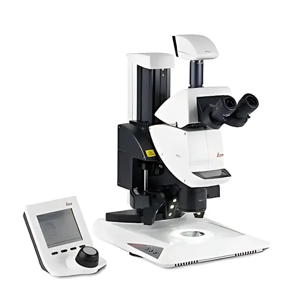
Leica M205 Stereo Microscope
| Brand | Leica |
|---|---|
| Origin | Germany |
| Model | M205 |
| Total Magnification | 1280× |
| Zoom Ratio | 7.8×–160× |
| Working Distance | 61.5 mm (with 1× Plan Apochromat objective) |
| Objective Type | Plan Apochromat |
| Illumination | LED5000RL Ring Light |
| Field of View | Ø59 mm (max), 1.44 mm (min) |
| Numerical Aperture | 0.35 |
| Resolution | 1050 lp/mm (max, with optimized optical configuration) |
| Viewing Angle | 30° trinocular tube |
| FusionOptics™ Technology | Standard (M205C/M205A) |
| Depth of Field Optimization | Dual-channel independent depth/resolution balancing |
| Encoding | Motorized or manual magnification encoding (model-dependent) |
| Optical Architecture | Common Main Objective (CMO), parallel beam path, lead-free housing (<2 s discharge time, surface resistivity 2×10¹¹ Ω/mm²) |
Overview
The Leica M205 Stereo Microscope is a high-performance CMO (Common Main Objective) architecture stereomicroscope engineered for demanding research, quality control, and precision manufacturing applications. Utilizing a dual-path optical design with a shared high-NA main objective, the M205 delivers exceptional resolution, color fidelity, and depth perception across its full zoom range. Its core innovation—FusionOptics™—employs asymmetric optical pathways: the right channel prioritizes spatial resolution (up to 1050 line pairs per millimeter), while the left channel maximizes depth of field. The human visual system fuses these complementary inputs, enabling simultaneous perception of fine structural detail and three-dimensional context—a capability unattainable with conventional stereo optics. Designed and manufactured in Wetzlar, Germany, the instrument complies with ISO 10934-1 (optical microscopy terminology), DIN EN ISO 9001 quality management standards, and incorporates RoHS-compliant, lead-free optical housing materials with electrostatic-dissipative surfaces (2×10¹¹ Ω/mm², <2 s decay from 1000 V to 100 V).
Key Features
- FusionOptics™ technology enables real-time, brain-fused visualization of high-resolution detail and extended depth of field without mechanical compromise
- Zoom ratio of 7.8×–160× (20.5:1 mechanical zoom range) supports seamless transition from macro overview to sub-micron inspection
- Plan apochromat objectives correct chromatic and spherical aberrations across visible and near-visible spectra; field number 23 ensures wide-field overview imaging
- Standard 30° inclined trinocular tube with 5°–45° adjustable viewing angle and 20-step diopter compensation ensures ergonomic adaptability for extended use
- Integrated LED5000RL ring illumination provides uniform, shadow-free, cool-white (5000 K) illumination controllable via hardware interface or Leica Application Suite (LAS)
- Motorized or manually encoded magnification systems (M205A vs. M205C) enable repeatable, audit-trail-capable configuration recall—critical for GLP/GMP environments
- Modular CMO platform supports interchangeable objectives (0.5×–2×), stands, digital cameras, and specialized lighting modules without optical recalibration
Sample Compatibility & Compliance
The Leica M205 accommodates specimens ranging from whole biological organisms (e.g., zebrafish embryos, insect dissections) to microelectronic assemblies, semiconductor wafers, and precision machined components. Its maximum working distance of 135 mm (with 0.5× objective) and minimum of 20.1 mm (with 2× objective) allows flexible integration with micromanipulators, soldering stations, and automated sample handlers. All optical components meet ISO 10110 surface quality standards; apochromatic correction satisfies ISO 10934-2 requirements for chromatic fidelity. The system supports compliance with FDA 21 CFR Part 11 when used with LAS X software configured for electronic signatures, audit trails, and user-access-controlled method locking.
Software & Data Management
Leica Application Suite (LAS) X provides full instrument control—including motorized zoom, focus, illumination intensity, and objective turret positioning—as well as image acquisition, annotation, measurement (ISO 13565-3 compliant), and report generation. Configuration data (magnification, objective ID, illumination mode) is automatically embedded into TIFF and JPEG metadata. LAS X supports DICOM export for pathology workflows and HDF5 for quantitative 3D reconstruction. Encoded hardware enables method-based parameter recall: up to 14 preset configurations can be stored and reloaded on M205C units; M205A models support unlimited cloud-synced protocols. All settings are timestamped and user-attributed, satisfying traceability requirements under ISO/IEC 17025 and GxP frameworks.
Applications
- Electronics manufacturing: SMT component inspection, PCB solder joint analysis, wire bonding validation
- Life sciences: Embryology, microdissection, Drosophila genetics, histological slide screening
- Materials science: Fracture surface analysis, coating thickness assessment, particle morphology classification
- Forensics: Toolmark comparison, fiber identification, gunshot residue documentation
- Quality assurance: Automotive component verification, medical device assembly checks, additive manufacturing layer inspection
FAQ
What distinguishes FusionOptics™ from conventional stereo microscopy?
FusionOptics™ uses two optically independent pathways—one optimized for resolution, the other for depth—fused neurologically by the observer. This eliminates the traditional trade-off between sharpness and depth perception inherent in single-path systems.
Is the M205 compatible with third-party digital cameras?
Yes, via standardized C-mount and F-mount adapters; however, full encoding and LAS integration require Leica-certified cameras (e.g., DFC9000 GT, ICC50 HD).
Can the M205 meet regulatory requirements for pharmaceutical QC labs?
When deployed with LAS X in validated configuration and paired with encoded hardware, it supports 21 CFR Part 11 compliance for electronic records and signatures.
What is the maximum usable resolution at highest magnification?
At 1280× total magnification (with 2× objective and 10× eyepieces), theoretical resolution is ~476 nm (calculated per Abbe limit, λ = 550 nm, NA = 0.35), verified experimentally at 1050 lp/mm using USAF 1951 test targets.
Does the system support automated focus mapping for 3D surface reconstruction?
Yes—when combined with LAS X Z-stack module and motorized focus drive (M205A), it acquires calibrated Z-series stacks with sub-micron step resolution and generates height maps compliant with ISO 25178-2 surface texture parameters.


