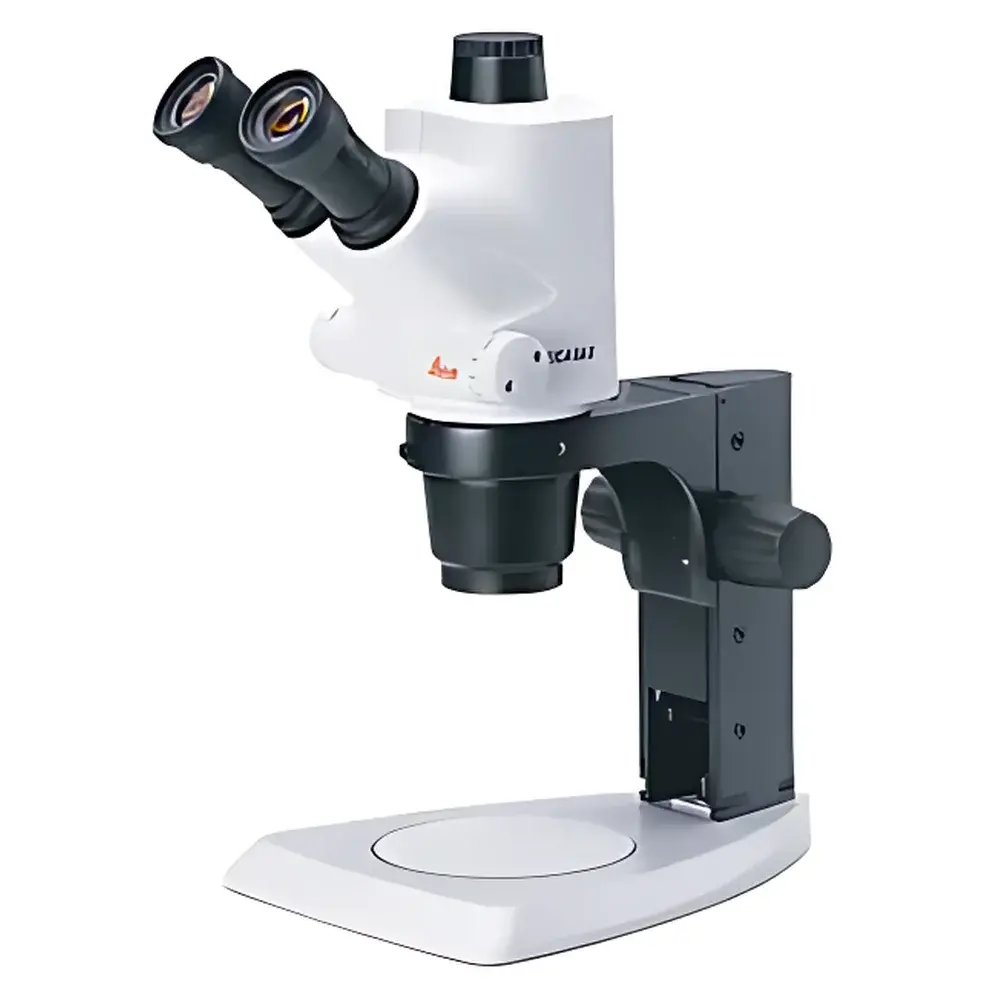
Leica S6 Stereo Microscope
| Brand | Leica |
|---|---|
| Origin | Germany |
| Model | S6 |
| Zoom Ratio | 6.3:1 |
| Magnification Range | 6.3× to 40× |
| Viewing Angle | 60° (S6 standard), 38° (S6 E variant) |
| Working Distance | 110 mm |
| Eyepiece Field of View | 23 mm (with 10× eyepieces), up to 36.5 mm maximum |
| Electrostatic Dissipation | 10⁶ Ω/square (S6 T variant) |
| Imaging Interface | 100% photo/video port with 100% light path toggle (S6 D variant) |
Overview
The Leica S6 Stereo Microscope is a high-performance, modular stereomicroscope engineered for precision visual inspection, quality control, and routine laboratory documentation across industrial, academic, and life science applications. Based on Greenough optical design principles, the S6 delivers true stereoscopic imaging with excellent depth perception, high contrast, and minimal chromatic aberration—ensuring accurate morphological assessment of three-dimensional specimens. Its 6.3:1 continuous zoom system provides a magnification range from 6.3× to 40×, enabling seamless transitions between macro- and meso-scale observation without mechanical stage repositioning. With a generous 110 mm working distance, the instrument accommodates bulky samples—including PCB assemblies, biological tissue blocks, or geological specimens—while maintaining ergonomic access for manipulation tools and ancillary instrumentation.
Key Features
- Optimized Greenough optics delivering high-resolution, distortion-free stereoscopic imaging with natural depth rendition.
- 6.3:1 zoom system with dual, user-definable mechanical limit stops—enabling repeatable magnification settings for standardized workflows.
- Ergonomic 60° inclined binocular viewing head (standard); optional 38° low-angle configuration (S6 E) for extended operator comfort during prolonged use.
- 10× widefield eyepieces providing a 23 mm field of view; compatible with optional 15× or 20× eyepieces to extend effective magnification while preserving image fidelity.
- Dedicated imaging variants: S6 D integrates a fully coupled photo/video port with 100% light path toggle—diverting all illumination to the camera sensor without compromising ocular image quality.
- S6 T variant features a conductive polymer housing with surface resistivity of 10⁶ Ω/square, meeting IEC 61340-5-1 requirements for electrostatic discharge (ESD)-sensitive environments such as semiconductor handling or microelectronics assembly.
Sample Compatibility & Compliance
The Leica S6 supports a broad spectrum of non-destructive sample types—from metallic components and solder joints to botanical sections, insect morphology, and forensic evidence. Its large working distance and open optical architecture accommodate accessories including stage micromanipulators, polarizing filters, transmitted light bases (e.g., Leica TL4000), and incident LED illuminators (e.g., Leica KL2500 LCD). The system complies with ISO 10934-1 (optical microscopy terminology), EN 61000-6-3 (EMC emissions), and RoHS 2011/65/EU directives. The S6 T model satisfies ANSI/ESD S20.20 and IEC 61340-5-1 for static-control workstations in cleanroom and electronics manufacturing settings.
Software & Data Management
When paired with Leica Application Suite (LAS X) software, the S6 D enables full digital workflow integration—including live image annotation, measurement calibration (ISO 10934-2 compliant), multi-focus stacking, and automated report generation. LAS X supports FDA 21 CFR Part 11-compliant audit trails, electronic signatures, and role-based user permissions—facilitating GLP and GMP-aligned documentation in regulated laboratories. Image metadata (magnification, illumination mode, timestamp, operator ID) is embedded directly into TIFF or JPEG files, ensuring traceability and data integrity across QA/QC records.
Applications
- Electronics manufacturing: Visual inspection of solder joints, component placement, and PCB trace continuity under variable magnification.
- Life sciences: Dissection support, embryo screening, and histological slide triage with simultaneous binocular viewing and digital capture.
- Materials science: Surface defect analysis, coating uniformity assessment, and particle morphology evaluation.
- Forensics: Toolmark comparison, fiber identification, and document examination requiring depth-resolved visualization.
- Education: Modular configuration allows curriculum-aligned adaptation—from introductory anatomy labs to advanced microfabrication training.
FAQ
What is the maximum field of view achievable with the Leica S6?
With 10× widefield eyepieces and the lowest zoom setting (6.3×), the field of view measures 36.5 mm in diameter.
Can the Leica S6 be integrated with third-party cameras?
Yes—the S6 D variant includes a C-mount adapter and standardized 1″ photo port, supporting industry-standard CCD/CMOS sensors and machine vision cameras.
Is the S6 compatible with transmitted light illumination?
Yes—optional transmitted light bases (e.g., Leica TL4000) can be mounted beneath the stage for transparent or semi-transparent specimen observation.
Does the S6 meet regulatory requirements for pharmaceutical QC environments?
While the base microscope is not intrinsically classified for GMP production areas, its S6 D configuration—when used with LAS X software—supports 21 CFR Part 11 compliance for electronic recordkeeping in regulated quality assurance processes.
How is electrostatic protection implemented in the S6 T model?
The S6 T incorporates a conductive polymer housing with surface resistivity of 10⁶ Ω/square, dissipating static charge at ≥10⁶ Ω/sq per IEC 61340-5-1, making it suitable for ESD-sensitive microelectronic handling.


