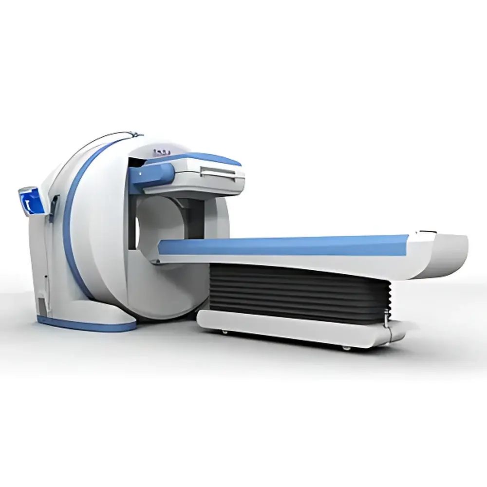
Hamamatsu BHP6601 Single-Head SPECT System
| Brand | Hamamatsu Photonics (BHP) |
|---|---|
| Model | BHP6601 |
| Type | Single-Head Gamma Camera-Based SPECT System |
| Effective Field of View | ≥510 mm × 390 mm |
| Energy Range | 50–400 keV |
| Intrinsic Spatial Resolution | ≤3.8 mm FWHM |
| Maximum Count Rate | ≥140 kcps |
| Intrinsic Uniformity (Differential) | ≤2.2% |
| (Integral) | ≤2.8% |
| Dimensions (System) | 170 × 183 × 200 cm |
| Weight (System) | 1950 kg |
| Patient Table | 266 × 57 × 65 cm, 250 kg |
| Power Cabinet | 43 × 50 × 96 cm, 47 kg |
| Collimator Cart (Single) | 71 × 83 × 124 cm, 180 kg |
| Software | Full-featured, Chinese-language native interface |
Overview
The Hamamatsu BHP6601 is a single-head gamma camera-based SPECT (Single Photon Emission Computed Tomography) system engineered for clinical nuclear medicine departments seeking a robust, cost-optimized entry point into functional molecular imaging. Designed around a high-sensitivity NaI(Tl) scintillation detector coupled with a parallel-hole collimator architecture, the BHP6601 operates on the principle of gamma photon detection following radiopharmaceutical administration (e.g., 99mTc, 123I, 131I, 67Ga). Its mechanical gantry rotates continuously or step-and-shoot to acquire projection data over 180° or 360°, which is reconstructed using filtered back-projection or iterative algorithms (OSEM) to generate transaxial, coronal, and sagittal tomographic slices. Unlike hybrid PET/CT or dual-head SPECT systems, the BHP6601 prioritizes operational simplicity, serviceability, and regulatory adaptability—making it particularly suitable for hospitals initiating or expanding nuclear medicine services under constrained capital budgets and staffing capacity.
Key Features
- Optimized single-head configuration delivering full clinical SPECT capability—including gated cardiac, dynamic renal, and brain perfusion studies—without requiring dual-detector infrastructure or elevated shielding requirements.
- Large effective field of view (≥510 mm × 390 mm) supporting whole-body bone scans and extended anatomical coverage in a single acquisition.
- Broad energy acceptance (50–400 keV) enabling compatibility with major diagnostic radionuclides used in routine nuclear medicine practice.
- Intrinsic spatial resolution ≤3.8 mm FWHM at center-of-field ensures adequate detail for myocardial wall definition, thyroid nodule characterization, and hepatic lesion localization.
- High intrinsic count rate capability (≥140 kcps) minimizes dead-time losses during high-activity injections, preserving quantitative accuracy in dynamic protocols such as renal DTPA or hepatobiliary HIDA studies.
- DICOM 3.0–compliant image export and network interface allow seamless integration into hospital-wide PACS and RIS environments, supporting standardized workflow and long-term archiving.
- Native Chinese-language acquisition and reconstruction software reduces training overhead and supports rapid protocol deployment by technologists without advanced English proficiency.
Sample Compatibility & Compliance
The BHP6601 accommodates standard clinical radiopharmaceuticals labeled with gamma-emitting isotopes approved for human use under national regulatory frameworks (e.g., NMPA Class III registration in China). It supports all common SPECT radiotracers including 99mTc-MDP (bone), 99mTc-sestamibi (myocardial perfusion), 99mTc-DTPA (renal dynamics), 99mTc-HIDA (hepatobiliary), 123I-NaI (thyroid), and 99mTc-RBC (gated blood pool). System calibration, uniformity verification, and center-of-rotation alignment follow AAPM Report No. 23 and IEC 61675-1 standards. All software modules comply with DICOM PS3.3 (Information Object Definitions) and PS3.4 (Service Classes), ensuring interoperability with third-party viewing and reporting platforms. While not FDA-cleared or CE-marked out-of-the-box, the system architecture supports documentation packages required for local regulatory submission and GLP-aligned QA recordkeeping.
Software & Data Management
The embedded acquisition and processing suite provides complete workflow control—from patient registration and protocol selection to reconstruction, quantification, and report generation. Reconstruction options include ramp-filtered back-projection and ordered-subset expectation maximization (OSEM) with attenuation correction (using uniform or Chang-based models) and scatter correction (dual-energy window method). Quantitative tools support time-activity curve analysis, eGFR estimation from renogram data, left ventricular ejection fraction (LVEF) calculation via gated SPECT, and semi-quantitative uptake ratios (e.g., thyroid uptake %). All images and metadata are stored in DICOM format with embedded modality-specific tags (e.g., SPECT Image Storage SOP Class UID). Audit trails log user actions, parameter changes, and reconstruction settings—supporting traceability requirements under ISO 13485 and national medical device quality management regulations.
Applications
- Myocardial Perfusion Imaging: Stress/rest 99mTc-sestamibi or tetrofosmin SPECT for ischemia detection and viability assessment.
- Whole-Body Bone Scintigraphy: Detection of metastatic disease, Paget’s disease, and occult fractures using 99mTc-MDP.
- Thyroid Imaging & Uptake: Static planar and SPECT/CT fusion-ready imaging of 123I or 99mTc-pertechnetate distribution; quantitative uptake measurement.
- Renal Dynamic Studies: Time-resolved evaluation of glomerular filtration rate (GFR), split renal function, and urinary obstruction with 99mTc-DTPA or 99mTc-MAG3.
- Cerebral Perfusion SPECT: Assessment of regional cerebral blood flow in epilepsy, dementia, and cerebrovascular disease using 99mTc-HMPAO or 99mTc-ECD.
- Hepatobiliary Imaging: Evaluation of biliary anatomy, gallbladder ejection fraction, and sphincter of Oddi dysfunction with 99mTc-HIDA.
- Lung Ventilation/Perfusion (V/Q) Scanning: Diagnosis of pulmonary embolism using 99mTc-MAA (perfusion) and 99mTc-DTPA aerosol (ventilation).
FAQ
Is the BHP6601 compliant with international imaging standards such as DICOM and IHE?
Yes—the system fully implements DICOM 3.0 Part 1–18 and supports key IHE Radiology Technical Framework actors including Modality Worklist (MWL), Modality Performed Procedure Step (MPPS), and Storage Commitment.
Can the BHP6601 perform quantitative SPECT (qSPECT)?
It supports semi-quantitative analysis (e.g., uptake ratios, time-activity curves) and includes attenuation/scatter correction tools required for basic qSPECT workflows, though absolute quantification requires additional phantom-based calibration and vendor-specific dose calibrator integration.
What maintenance and service support does Hamamatsu provide for this system?
Hamamatsu Photonics offers factory-trained field service engineers, remote diagnostics support, annual preventive maintenance contracts, and guaranteed spare parts availability for ≥10 years post-installation.
Does the system support integration with hospital EMR or RIS platforms?
Yes—via HL7 v2.x ADT and ORU messages, enabling automatic patient demographic import and structured report export to EMR systems.
Is the software upgradable to support future SPECT/CT hybrid functionality?
The BHP6601 is a dedicated SPECT-only platform; CT integration is not supported. For hybrid configurations, Hamamatsu recommends the STS series or third-party CT co-registration solutions.


