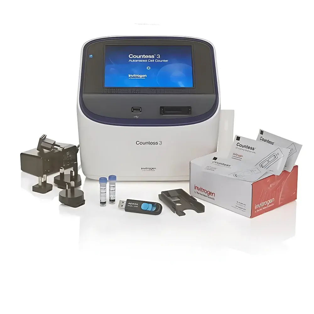
Thermo Scientific Invitrogen Countess 3 and Countess 3 FL Automated Cell Counters
| Brand | Thermo Scientific |
|---|---|
| Origin | Singapore |
| Model Numbers | A49891, A49893 |
| Measurement Time | 10 s/sample |
| Cell Concentration Range | 1×10⁴–1×10⁷ cells/mL |
| Sample Volume | 0.4 µL |
| Throughput | 2 samples |
| Detectable Cell Size | 4–60 µm |
Overview
The Thermo Scientific Invitrogen Countess 3 and Countess 3 FL Automated Cell Counters are benchtop digital imaging systems engineered for rapid, reproducible, and objective quantification of mammalian, insect, yeast, and bacterial cells in suspension. These instruments employ high-resolution brightfield microscopy coupled with AI-driven image analysis—specifically, a deep learning neural network trained on thousands of annotated cell images—to distinguish viable from non-viable cells (when stained with trypan blue or similar dyes), resolve clustered cells, suppress debris interference, and deliver statistically robust counts without user-dependent thresholding or manual gating. Unlike conventional hemocytometer-based methods, the Countess 3 platform eliminates inter-operator variability by automating focus, illumination intensity, and image acquisition—ensuring consistent measurement conditions across users, shifts, and timepoints. The system operates on the principle of morphometric segmentation: raw grayscale images are processed through convolutional neural networks (CNNs) that classify pixel clusters based on learned features—including edge contrast, circularity, size distribution, and staining homogeneity—enabling accurate enumeration even for challenging samples such as RAW 264.7 macrophages, rodent PBMCs, and aggregated primary neurons.
Key Features
- AI-Powered Cell Recognition: Proprietary deep learning algorithm trained on diverse cell types and morphologies to differentiate intact cells from clumps and debris; validated for accuracy across >20 common cell lines and primary isolates.
- Automated Imaging Workflow: Motorized stage, auto-focus, and dynamic LED illumination adjustment ensure optimal image capture without manual intervention—reducing operator training burden and measurement drift.
- Integrated Calculators: Onboard pre-dilution and passaging calculators use real-time count data to compute required diluent volumes, seeding densities, and media volumes—supporting GLP-aligned documentation workflows.
- Multi-Format Reporting: Export results as PNG, JPG, TIFF, PDF, CSV, or FCS (Flow Cytometry Standard) files—enabling traceability, LIMS integration, and downstream analysis in third-party software.
- Reusable Slide Compatibility: Designed exclusively for Invitrogen Countess reusable slides (Cat. No. AMQAF1000), minimizing consumable cost per sample and reducing plastic waste versus disposable alternatives.
Sample Compatibility & Compliance
The Countess 3 series supports a broad range of suspension-cultured cells—including adherent cells detached via enzymatic or non-enzymatic methods—as well as yeast (e.g., Saccharomyces cerevisiae), bacteria (e.g., E. coli DH5α), and algae. Viability assessment is compatible with standard membrane-impermeant dyes including trypan blue, propidium iodide (PI), and fluorescein diacetate (FDA). The Countess 3 FL extends functionality with dual interchangeable fluorescence channels (user-installed EVOS™ light cubes), supporting excitation/emission pairs from DAPI (358/461 nm) to Cy5 (649/670 nm), enabling simultaneous two-color viability assays (e.g., PI + Hoechst 33342) or transfection efficiency quantification (e.g., GFP + RFP). All instruments comply with IEC 61010-1 safety standards and support audit-ready operation under FDA 21 CFR Part 11 when deployed with appropriate IT infrastructure and electronic signature protocols.
Software & Data Management
The Countess 3 Control Software (v3.x) provides a locked-down, single-purpose interface optimized for cell culture labs—eliminating unnecessary functions that could compromise data integrity. Each measurement generates a timestamped metadata log containing instrument ID, slide serial number, user login (if enabled), exposure settings, and raw image hash—facilitating full traceability per ISO/IEC 17025 and GLP requirements. Data export supports CSV for Excel-based QC trending, FCS for compatibility with flow cytometry analysis platforms (e.g., FlowJo, FCS Express), and PDF reports with embedded histograms and gate overlays. Optional network connectivity allows centralized data archiving via SMB or FTP, while local storage retains ≥1,000 measurement records with automatic overwrite protection.
Applications
- Quantitative monitoring of cell proliferation kinetics during passage expansion
- Viability assessment pre- and post-cryopreservation or transfection
- QC release testing of bioproduction seed stocks (e.g., CHO, HEK293)
- Standardization of cell seeding density for assay development (e.g., ELISA, cytotoxicity, CRISPR screening)
- Fluorescent reporter validation in stable or transient expression systems
- Microbial load estimation in fermentation or probiotic manufacturing
FAQ
What is the minimum required sample volume?
0.4 µL per measurement—loaded onto the reusable slide’s dual chambers (2 samples per slide).
Can the Countess 3 perform viability analysis without fluorescent dyes?
Yes—the Countess 3 (brightfield-only) uses trypan blue exclusion; the Countess 3 FL adds fluorescent viability dyes (e.g., PI, 7-AAD) for enhanced specificity in heterogeneous populations.
Is the AI algorithm customizable or trainable with user-specific cell types?
No—the neural network is factory-trained and locked to ensure regulatory consistency; however, its generalization performance has been verified across >50 cell types in independent validation studies.
How does the system handle cell clumping during counting?
The deep learning model identifies internal texture gradients and edge discontinuities within aggregates, enabling sub-cluster segmentation and individual cell enumeration—validated against manual counts using calibrated microbeads and dissociated spheroids.
Are software updates provided, and how are they installed?
Firmware and software updates are distributed free-of-charge via Thermo Fisher Connect; installation requires USB drive transfer and guided in-app wizard—no internet connection needed during operation.
Related Products


