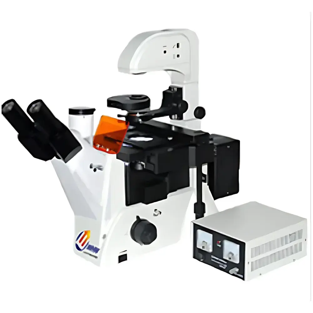
YANRUN FM-200 Inverted Infinity-Corrected Phase Contrast & Epi-Fluorescence Microscope
| Brand | YANRUN |
|---|---|
| Origin | Shanghai, China |
| Manufacturer Type | Direct Manufacturer |
| Model | FM-200 Inverted Infinity-Corrected Phase Contrast & Epi-Fluorescence Microscope |
| Optical Magnification | 100×–400× |
| Eyepieces | Widefield WF10X (Φ22 mm), Centering Telescope |
| Objective Lenses | Infinity-Corrected Achromatic PLL (Cover Glass Thickness 1.2 mm): 10×/0.25 (WD 4.3 mm), 20×/0.4 (WD 8.0 mm), 40×/0.6 (WD 3.5 mm) |
| Phase Contrast PLL/PHP2 Objectives | Same Specifications |
| Trinocular Head | 45° Inclination, 100% Light Path Diversion for Projection, Interpupillary Distance 53–75 mm |
| Transmitted Illumination | 6 V / 30 W Halogen Lamp with Dimming Control |
| Phase Contrast Condenser | Rotating Carousel-Type, WD 55 mm |
| Filter Set | Blue, Green, Ground Glass |
| Epi-Fluorescence Illumination | 100 W Mercury Arc Lamp, Dual-Voltage Power Supply (110 V / 230 V) |
| Excitation Filter Sets | Blue-Green Set (Ex: 450–490 nm / Em: ≥515 nm |
| Ex | 495–555 nm / Em: ≥595 nm) |
| UV-Violet Set (Ex | 320–380 nm / Em: ≥435 nm |
| Ex | 380–415 nm / Em: ≥475 nm) |
| Stage Dimensions | 208 × 227 mm, Travel Range 77 × 114 mm |
| Circular Glass Stage | Φ118 mm |
| Nosepiece | Quintuple Revolving |
| Specimen Holders | Three Standard Culture Dish Adapters (for Ø87.5 mm, Ø68.5 mm, and Rectangular 57 × 82 mm Dishes) |
| Focus Mechanism | Coaxial Coarse/Fine Adjustment with Locking & Upper/Lower Limits, Fine Focus Graduation: 2 µm per Division |
| Optional Accessories | Graticule Eyepieces (0.1 mm/div), 5×/0.12 Objective, Hexaposition Nosepiece, Custom Dish Adapters (T25, 35 mm, 50 mm, 66 × 66 mm, Slide Holder), CCD Adapters (0.4×, 0.5×, 1×, 0.5× with Graticule), USB/VIDEO & Digital Camera Interfaces |
Overview
The YANRUN FM-200 is an inverted infinity-corrected microscope engineered for live-cell imaging in biological and biomedical research laboratories. Its optical architecture integrates phase contrast and epi-fluorescence modalities within a single robust platform, enabling high-contrast observation of unstained, transparent specimens—such as adherent or suspension-cultured cells—without fixation or labeling. The system employs Köhler illumination for both transmitted and reflected light paths, ensuring uniform intensity distribution and optimal resolution across the full field of view. Designed around an inverted configuration, the FM-200 positions objectives beneath the specimen stage while locating condensers and light sources above—ideal for long-term time-lapse studies in standard tissue culture vessels. All objective lenses are infinity-corrected achromats with standardized parfocal distance (45 mm) and cover-glass correction (1.2 mm), supporting consistent image quality across magnifications from 100× to 400×.
Key Features
- Hinged trinocular head inclined at 45°, providing ergonomic viewing and 100% light path diversion for simultaneous visual observation and digital imaging.
- Large-format mechanical stage (208 × 227 mm) with 77 × 114 mm travel range and three interchangeable culture dish adapters—supporting common formats including Ø68.5 mm, Ø87.5 mm, and rectangular dishes up to 57 × 82 mm.
- Rotating, swing-in/swing-out condenser assembly with 55 mm working distance—enabling unobstructed access to tall culture flasks and cylindrical bioreactors without contamination risk.
- Dual-mode illumination: adjustable 6 V / 30 W halogen lamp for brightfield and phase contrast; independently controlled 100 W mercury arc lamp with dual-voltage power supply (110 V / 230 V) for stable epi-fluorescence excitation.
- Four pre-aligned fluorescence filter sets: blue/green (FITC/TRITC-equivalent), ultraviolet, and violet—each with certified excitation/emission bandpass characteristics compliant with ISO 10934-1 spectral tolerances.
- Coaxial coarse/fine focusing mechanism with mechanical stop limits and 2 µm fine-focus graduation—optimized for precise Z-stack acquisition in confocal-compatible workflows.
Sample Compatibility & Compliance
The FM-200 accommodates standard in vitro cell culture formats—including Petri dishes, multi-well plates, T-flasks, and custom bioreactor chambers—via its modular stage insert system. The transparent stage plate permits real-time verification of objective engagement during nosepiece rotation, minimizing accidental collisions. All optical components meet ISO 8578 (microscope mechanical tube length and parfocal distance) and ISO 10934-1 (fluorescence filter spectral performance) requirements. While not FDA-cleared as a diagnostic device, the system supports GLP-compliant documentation when paired with audit-trail-enabled imaging software (e.g., via optional USB/VIDEO interface with timestamped metadata embedding). Phase contrast optics conform to ISO 8578 Annex B for quantitative contrast transfer function (CTF) validation.
Software & Data Management
The FM-200 features a rear-mounted C-mount interface (standard 1×) compatible with industrial-grade CMOS/CCD cameras and scientific-grade sCMOS sensors. Optional adapters—including 0.4×, 0.5×, and 1× reduction optics with calibrated graticules (0.1 mm/div)—enable pixel-to-micron calibration traceable to NIST standards. When integrated with third-party acquisition platforms (e.g., MicroManager, NIS-Elements, or HALO Imaging Suite), the microscope supports automated multi-channel fluorescence capture, Z-stacking, time-lapse scheduling, and metadata tagging aligned with MIAME and OME-TIFF specifications. Image export formats include TIFF (16-bit linear), PNG, and JPEG2000—preserving dynamic range for downstream quantitative analysis in MATLAB, Python (OpenCV, scikit-image), or ImageJ/Fiji.
Applications
- Long-term monitoring of stem cell differentiation and organoid development in physiological culture environments.
- Real-time assessment of transfection efficiency using GFP/RFP-tagged constructs under blue/green excitation.
- Quantitative phase contrast analysis of red blood cell morphology and membrane fluctuations in hematology research.
- UV-excited DAPI staining for nuclear segmentation and mitotic index determination in cancer biology assays.
- High-content screening in 96-well plates using motorized XY-stage add-ons (available separately).
- Teaching laboratory use for foundational instruction in cell biology, microbiology, and histology techniques.
FAQ
Is the FM-200 suitable for quantitative fluorescence intensity measurements?
Yes—when used with calibrated cameras and reference standards (e.g., fluorescent microspheres traceable to NIST SRM 2801), the system supports semi-quantitative intensity profiling across multiple channels.
Can I upgrade to motorized focus or stage control?
The FM-200’s mechanical architecture supports aftermarket integration of stepper-motorized Z-drives and XY translation stages via standard M4 mounting threads and TTL trigger interfaces.
Does the mercury lamp require warm-up or cool-down cycles?
Yes—the 100 W Hg lamp requires a minimum 15-minute warm-up for spectral stabilization and a mandatory 10-minute post-use cooling period before restart to prevent arc tube degradation.
Are replacement filter sets available for other fluorophores (e.g., Cy5, Alexa Fluor 647)?
Custom excitation/emission filter sets can be ordered separately; compatibility must be verified against the lamp’s spectral output and the system’s dichroic mirror cut-on wavelengths.
What is the maximum recommended immersion depth for objective front lenses during live imaging?
All standard objectives are designed for dry use only; oil or water immersion variants are not supported in the base FM-200 configuration but may be added via custom OEM collaboration.


