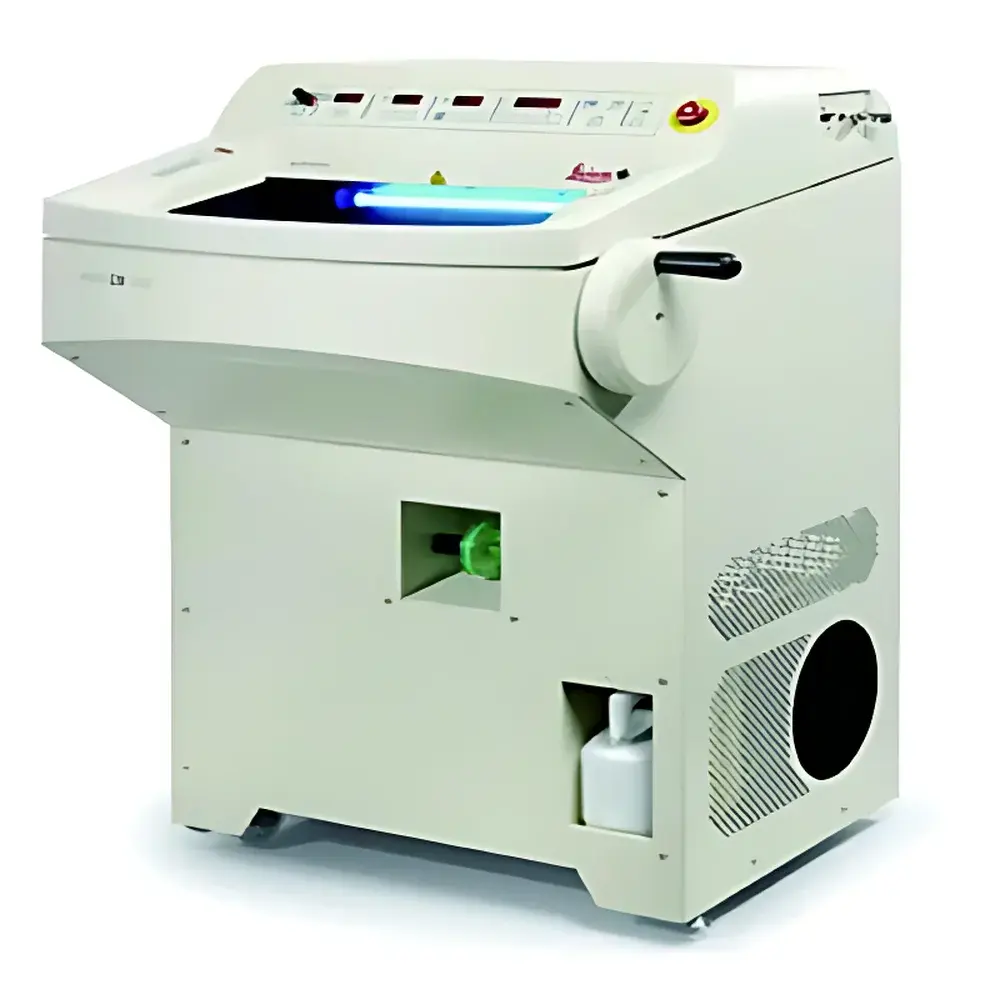
Leica CM1850 Cryostat Microtome
| Brand | Leica |
|---|---|
| Origin | Germany |
| Model | CM1850 |
| Type | Cryostat Microtome |
| Compliance | CE, ISO 13485 (Medical Device Quality Management), IEC 61010-1 (Safety Requirements for Laboratory Equipment) |
| Cooling System | Dual-stage Peltier cooling with rapid pre-cooling mode |
| Operating Temperature Range | –35 °C to –15 °C (adjustable in 1 °C increments) |
| Section Thickness Range | 0.5–100 µm (1 µm incremental adjustment) |
| Sample Stage Travel | X: ±20 mm, Y: ±15 mm, Z: 40 mm (motorized vertical sample lift) |
| Knife Holder | Universal tungsten carbide or stainless-steel knife compatibility |
| Safety Features | Interlocked cryochamber door, automatic blade guard activation, condensation management system |
| Dimensions (W×D×H) | 720 × 790 × 680 mm |
| Weight | 125 kg |
Overview
The Leica CM1850 Cryostat Microtome is a precision-engineered, CE-marked cryogenic microtome designed for high-fidelity frozen sectioning in diagnostic histopathology, neuroscience research, and translational biomedical laboratories. It operates on the fundamental principle of cryosectioning—rapidly freezing biological specimens to preserve native morphology and antigenicity, then cutting thin, consistent sections at sub-zero temperatures using a motorized rotary mechanism and thermally stabilized specimen stage. Unlike room-temperature microtomes, the CM1850 integrates a dual-stage Peltier-based cooling architecture that achieves stable chamber temperatures between –35 °C and –15 °C, minimizing ice crystal artifact formation while enabling reproducible sectioning of soft, lipid-rich, or delicate tissues—including brain, adipose, and unfixed biopsy specimens. Its design adheres to IEC 61010-1 safety standards for laboratory equipment and is manufactured under Leica Microsystems’ ISO 13485-certified quality system, ensuring traceability and consistency across global clinical and research deployments.
Key Features
- Motorized, stepless section thickness control (0.5–100 µm) with 1 µm resolution, calibrated via integrated optical encoder for long-term repeatability
- Dual-zone thermal management: independent regulation of cryochamber ambient temperature and specimen block surface temperature to suppress thermal drift during serial sectioning
- Ergonomic, low-profile specimen stage with motorized Z-axis lift (40 mm travel) and fine-tuned X/Y positioning (±20 mm / ±15 mm), reducing operator fatigue during extended session workloads
- Interlocked cryochamber door with fail-safe power cutoff and real-time condensation detection—prevents frost accumulation on optics and knife edges
- Universal knife holder accommodating both disposable blade cassettes and reusable tungsten carbide knives, supporting standardized blade geometry per ASTM E2907 (Standard Guide for Microtomy)
- Integrated LED illumination with adjustable intensity and shadow-free optical path, optimized for brightfield microscopy validation prior to staining
Sample Compatibility & Compliance
The CM1850 supports a broad spectrum of unembedded and OCT-embedded tissues—including formalin-fixed paraffin-processed (FFPE) resections requiring rapid intraoperative assessment, fresh-frozen tumor biopsies, murine brain coronal sections, and plant vascular tissue. All mechanical and thermal parameters are validated against ISO/IEC 17025-accredited test protocols for microtome performance verification. The instrument complies with EU Medical Device Regulation (MDR 2017/745) as a Class I non-invasive device when used in diagnostic pathology workflows. Data integrity features—including timestamped operation logs and user-access-controlled parameter locking—support GLP and CAP-accredited laboratory requirements. Optional integration with LIS/HIS systems enables audit-ready documentation aligned with CLIA and CAP checklist ANP.42000 (Frozen Section Quality Control).
Software & Data Management
While the CM1850 operates via an intuitive tactile control panel (no embedded OS), its operational parameters—including section thickness, chamber temperature, cutting speed, and user ID—are logged locally in non-volatile memory with ISO-compliant time stamping (UTC). Exportable CSV logs support retrospective QC analysis and root-cause investigation in CAP/FDA 21 CFR Part 11–aligned environments when paired with Leica’s optional LAS X Connect module. Firmware updates are delivered via encrypted USB interface with SHA-256 signature verification, maintaining integrity throughout the device lifecycle. All configuration changes require multi-level authentication (operator + supervisor PIN), satisfying GxP documentation traceability requirements.
Applications
- Intraoperative frozen section diagnosis in surgical pathology, meeting College of American Pathologists (CAP) turnaround time benchmarks (<20 min from specimen receipt to sign-out)
- Pre-analytical preparation for spatial transcriptomics and MALDI imaging mass spectrometry, where section integrity and minimal thaw artifacts are critical
- Serial section reconstruction for 3D ultrastructural analysis in electron microscopy pipelines
- Quality control of biobank-derived tissue archives, particularly for rare disease cohorts requiring morphological preservation over decades
- Training platform in histotechnology programs accredited by the National Accrediting Agency for Clinical Laboratory Sciences (NAACLS)
FAQ
What is the minimum achievable section thickness, and how is thickness accuracy verified?
The CM1850 achieves 0.5 µm nominal thickness with ±0.2 µm repeatability (per ISO 21534:2021 Annex B calibration protocol using certified polymer test blocks). Accuracy is confirmed via optical interferometry during factory acceptance testing.
Does the CM1850 support remote monitoring or integration with laboratory information systems?
Yes—via optional LAS X Connect hardware, enabling HL7-compatible status reporting, error logging, and user activity auditing for compliance with 21 CFR Part 11 Subpart B.
Is routine maintenance documented and traceable for regulatory audits?
All service events—including Peltier module recalibration, knife alignment verification, and chamber seal integrity checks—are recorded in the onboard log with digital signature fields for technician and QA reviewer.
Can the CM1850 be used for cutting hard-tissue samples such as calcified bone or decalcified specimens?
No—the CM1850 is engineered exclusively for soft-to-medium consistency frozen tissues. Hard-tissue sectioning requires a dedicated sledge microtome (e.g., Leica SM2010 R) or diamond-knife ultramicrotome.
What safety certifications apply to the CM1850 in North American laboratories?
It carries cULus listing per UL 61010-1 and complies with ANSI Z358.1 for emergency eyewash proximity requirements when installed per Leica’s site planning guide (document ref. CM1850-SPG-EN v3.2).


