Yoke XSP-8CA Dual-Eyepiece Electrically Illuminated Biological Microscope
| Brand | Yoke |
|---|---|
| Origin | Shanghai, China |
| Manufacturer Type | Direct Manufacturer |
| Country of Origin | China |
| Model | XSP-8CA |
| Price Range | USD 280 – 700 |
| Eyepieces | Wide-Field WF10X, Huygens H16X |
| Objectives | Achromatic 4X, 10X, 40X(S), 100X(S) |
| Condenser | Abbe condenser with adjustable iris diaphragm and filter holder |
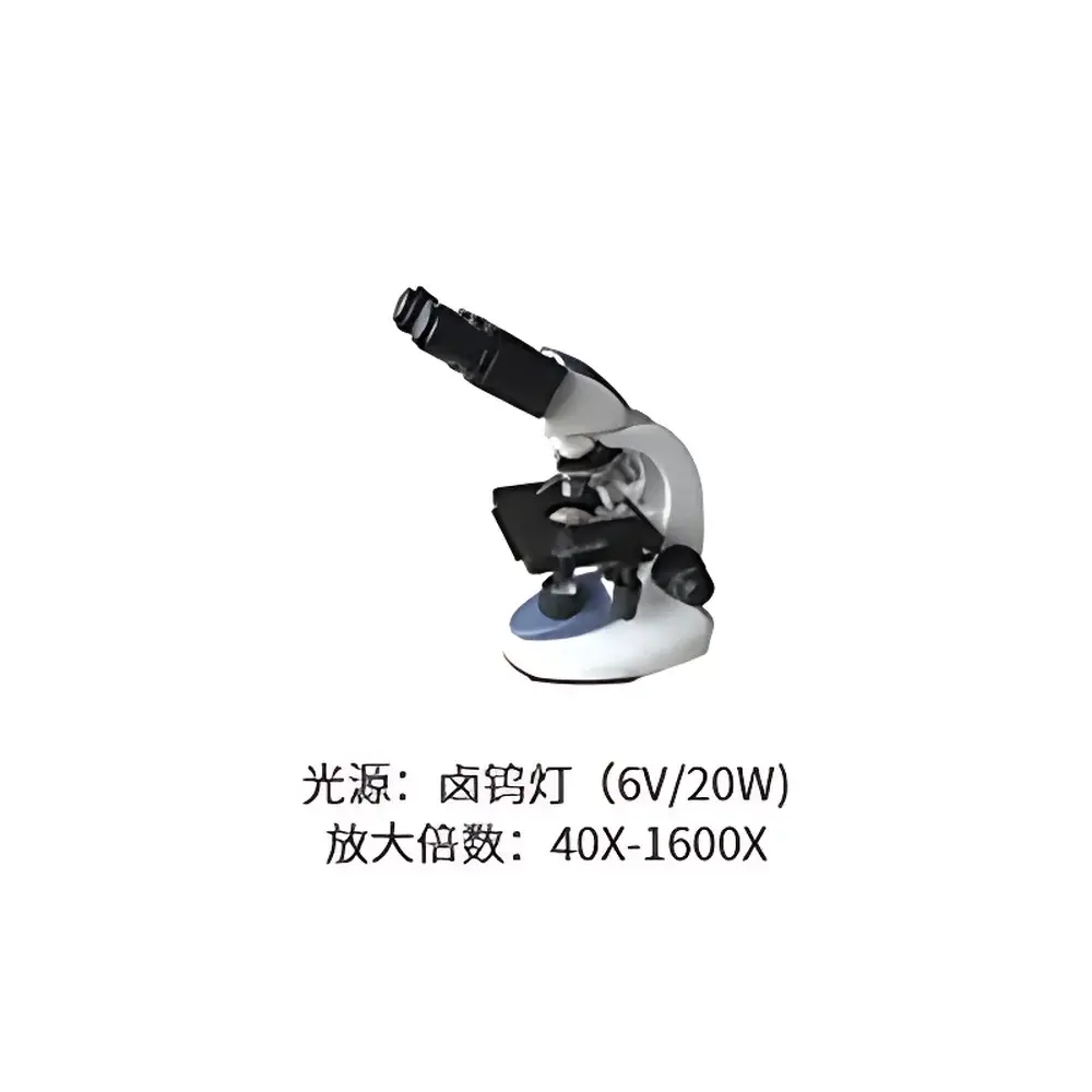
| Illumination | 6V/20W halogen lamp |
| Power Supply | AC-powered electrical illumination system |
Overview
The Yoke XSP-8CA is a dual-eyepiece, electrically illuminated biological microscope engineered for routine observation and instructional use in life science laboratories. It operates on conventional brightfield optical principles, utilizing Köhler illumination geometry to deliver uniform, glare-free specimen illumination across the field of view. Designed for fixed or live unstained and stained biological specimens—including blood smears, tissue sections, microbial cultures, and plant histological preparations—the instrument supports standard transmitted-light microscopy workflows in clinical diagnostics, academic teaching, and applied research settings. Its mechanical stage, coaxial coarse/fine focusing mechanism, and standardized DIN objective mounting ensure compatibility with widely adopted laboratory protocols and accessory ecosystems.
Key Features
- Dual-eyepiece optical head with 45° inclined tubes and interpupillary distance adjustment (55–75 mm), supporting extended user comfort during prolonged observation sessions.
- Achromatic objective lens set (4X, 10X, 40X spring-loaded, 100X oil immersion) meeting ISO 8578 tolerances for chromatic and spherical aberration correction, enabling reliable morphological assessment at magnifications up to 1600× (with H16X eyepiece).
- Abbe condenser (NA 1.25) with continuously adjustable iris diaphragm and integrated filter holder—optimized for contrast control and resolution enhancement under varying specimen thickness and staining density.
- Stable cast-metal frame with rack-and-pinion focusing system featuring 2 mm coarse travel and 0.002 mm fine-focus increment, ensuring precise Z-axis positioning for layered tissue analysis.
- 6V/20W halogen illumination module with integrated voltage regulation, delivering consistent color temperature (~3200 K) and luminous flux over extended operational cycles; compatible with standard 120/230 V AC input via external power adapter.
Sample Compatibility & Compliance
The XSP-8CA accommodates standard 1″ × 3″ glass microscope slides (thickness: 1.0–1.2 mm) and #0–#2 cover slips. It supports both dry and oil-immersion techniques, with the 100X objective requiring immersion oil (n = 1.515) for optimal numerical aperture utilization. The microscope conforms to ISO 10934-1:2001 (Microscopes — Nomenclature of parts) and meets mechanical safety requirements outlined in IEC 61010-1 for laboratory electrical equipment. While not certified for GLP/GMP-regulated production environments, its construction and calibration traceability support documentation-compliant use in educational labs, QC screening, and non-critical diagnostic support per CLIA-waived testing guidelines.
Software & Data Management
As a standalone optical instrument without integrated digital imaging hardware, the XSP-8CA does not include proprietary software or onboard data storage. However, it features a standardized trinocular port (optional accessory) enabling coupling with C-mount-compatible CCD or CMOS cameras (e.g., 1/2″ sensor format). When paired with third-party imaging platforms—such as Ocular Insight Suite, MicroManager (open-source), or HALCON-based acquisition systems—users can implement audit-trail-capable image capture, annotation, measurement (length, area, particle count), and export in TIFF, JPEG2000, or FITS formats. Metadata embedding (objective magnification, illumination setting, timestamp) complies with FAIR data principles for academic publication and internal lab record retention.
Applications
- Clinical hematology: Differential white blood cell counting, platelet morphology evaluation, and malaria parasite detection in Giemsa-stained thin films.
- Pharmaceutical microbiology: Sterility test verification, fungal hyphae characterization, and antibiotic susceptibility zone measurement (Kirby-Bauer).
- Plant and agricultural science: Pollen grain morphology, stomatal density quantification, and root nodule symbiont identification in legume studies.
- Undergraduate biology instruction: Mitosis staging in Allium root tip squashes, protozoan motility analysis, and comparative histology of vertebrate organ sections.
- Industrial quality control: Verification of cell culture monolayer confluence pre-harvest and detection of mycoplasma contamination in bioprocess monitoring workflows.
FAQ
Is the XSP-8CA compatible with fluorescence observation?
No—it lacks excitation filters, dichroic mirrors, and high-intensity LED or mercury vapor illumination required for epifluorescence. Upgrade paths require retrofitting with third-party fluorescence modules, which may compromise optical alignment integrity.
Can immersion oil be used with all high-magnification objectives?
Only the 100X (S) objective is designed for oil immersion; using oil with the 40X (S) objective may damage lens coatings and degrade image fidelity.
What is the maximum usable magnification without significant loss of resolution?
At 1000× total magnification (100X objective × 10X eyepiece), the system operates near its practical resolution limit (~0.2 µm under ideal Köhler conditions); higher nominal magnifications yield empty magnification without added structural detail.
Does the microscope support phase contrast or darkfield accessories?
Yes—its Abbe condenser accepts aftermarket phase contrast sliders and darkfield stops (e.g., BH2-series compatible units), though alignment validation is required post-installation.
Is service documentation available in English?
Yes—Yoke provides bilingual (Chinese/English) user manuals, optical alignment checklists, and maintenance schedules covering lamp replacement, dust mitigation, and mechanical recalibration procedures.
Related Products


