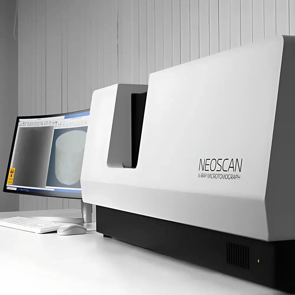
Neoscan N80 High-Resolution Benchtop Micro-CT System for Orthopedic Research
| Brand | Neoscan |
|---|---|
| Origin | Belgium |
| Detector Type | Flat-Panel Detector |
| Scan Mode | Sample Rotation Only (RO) |
| Spatial Resolution | 2 µm |
| X-ray Energy | 110 kV |
| Field of View | 100 mm × 163 mm |
| Maximum Sample Weight | 20 kg |
| Dimensions | 1200 mm × 640 mm × 520 mm |
| Precision | Micron-Level |
Overview
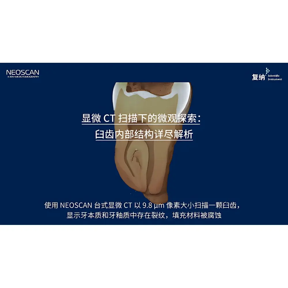
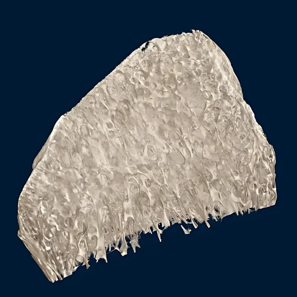
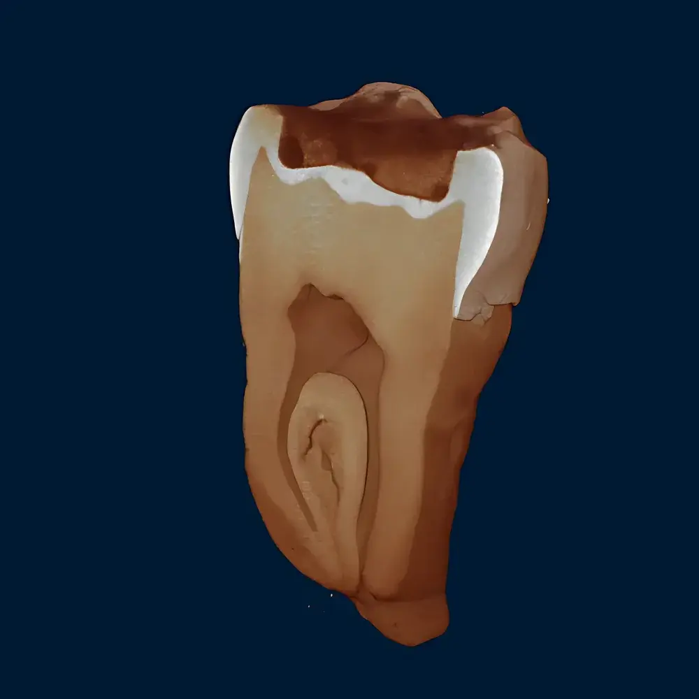
The Neoscan N80 High-Resolution Benchtop Micro-CT System is an engineered solution for non-destructive, high-fidelity 3D characterization of bone microarchitecture. Based on cone-beam X-ray computed tomography principles, it employs a fixed-source–fixed-detector geometry with precise sample rotation (RO mode), enabling quantitative volumetric imaging without physical sectioning. The system operates at 110 kV photon energy and achieves isotropic spatial resolution down to 2 µm—sufficient to resolve trabecular thickness, cortical porosity, lacunar-canalicular networks, and early-stage mineralization gradients in murine, ovine, and human bone specimens. Its benchtop footprint and robust mechanical architecture make it suitable for integration into university orthopedic labs, preclinical research facilities, and contract research organizations (CROs) conducting GLP-compliant bone morphometry studies.
Key Features
- Micron-level dimensional accuracy and repeatability across repeated scans, validated per ISO 12791-2 for micro-CT performance assessment
- Flat-panel detector with high dynamic range and low electronic noise, optimized for contrast discrimination in low-Z biological matrices
- Motorized precision rotation stage with sub-arcsecond angular positioning stability, minimizing motion-induced artifacts
- Integrated geometric calibration routine using certified reference phantoms (e.g., hydroxyapatite density standards, aluminum step wedges)
- Thermally stabilized X-ray source ensuring consistent beam hardening characteristics over extended acquisition sessions
- Benchtop form factor (1200 × 640 × 520 mm) designed for standard laboratory floor space and Class II biosafety cabinet compatibility
Sample Compatibility & Compliance
The N80 accommodates ex vivo bone samples up to 20 kg and 163 mm in height, supporting intact femora, vertebrae, tibiae, and large-format implant-bone interface specimens. It complies with ASTM E1441 (Standard Practice for Computed Tomography), ISO 15732 (Quantitative micro-CT for bone tissue), and supports traceable density calibration per NIST SRM 2197 (hydroxyapatite reference material). All raw projection data and reconstructed volumes are stored in DICOM 3.0 format, ensuring interoperability with PACS systems and third-party analysis platforms. The system architecture conforms to FDA 21 CFR Part 11 requirements for audit trails, electronic signatures, and data integrity when operated with optional validated software modules.
Software & Data Management
Acquisition and reconstruction are managed via Neoscan’s proprietary CT Studio Suite, which includes GPU-accelerated Feldkamp-Davis-Kress (FDK) reconstruction, ring artifact suppression, beam-hardening correction, and automatic center-of-rotation alignment. Post-processing tools support ISO/ASTM-standard bone morphometric analysis—including BV/TV, Tb.Th, Tb.Sp, Tb.N, Conn.D, SMW, and cortical thickness mapping—using calibrated grayscale-to-density conversion curves. Export formats include segmented STL for finite element modeling (FEM), NRRD for ImageJ/Fiji workflows, and HDF5 for Python-based machine learning pipelines. All processing steps are logged with timestamped metadata, supporting full traceability in regulatory submissions.
Applications
- Preclinical osteoporosis modeling: longitudinal quantification of trabecular degradation and cortical thinning in ovariectomized sheep or rodent models
- Orthopedic implant osseointegration: 3D assessment of bone-implant contact (BIC), gap formation, and peri-implant bone volume changes over time
- Fracture healing kinetics: temporal tracking of callus mineralization, bridging continuity, and remodeling progression at weekly intervals
- Biomaterial scaffold evaluation: pore interconnectivity analysis, degradation rate mapping, and mechanical property correlation via digital volume correlation (DVC)
- Cartilage-bone interface studies: co-registered segmentation of calcified cartilage, tidemark location, and subchondral plate porosity
FAQ
What is the minimum voxel size achievable under standard operating conditions?
The system achieves an effective isotropic voxel size of 2 µm at optimal magnification; actual voxel size depends on sample-to-detector distance and field-of-view selection.
Can the N80 perform in vivo scanning of small animals?
No—the N80 is configured exclusively for ex vivo specimen analysis. In vivo applications require dedicated preclinical imaging platforms with dose-reduction hardware and physiological gating.
Is the system compatible with third-party morphometric software such as BoneJ or CTAn?
Yes—reconstructed volumes are exported in standard 16-bit TIFF stacks or DICOM series, fully supported by BoneJ (ImageJ), CTAn (Bruker SkyScan), and MorphoGraphX.
How is geometric calibration maintained over time?
A daily automated calibration sequence uses a stainless-steel ball-bearing phantom; quarterly verification follows ISO 12791-2 procedures with traceable metrology-grade phantoms.
Does the system support automated batch scanning for multi-sample studies?
Yes—CT Studio includes scriptable acquisition protocols and queue management for unattended overnight runs, with real-time email alerts upon completion or error detection.


