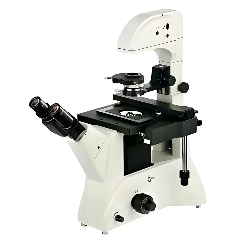
Betop Scientific XDS-3 Inverted Biological Microscope
| Brand | Betop Scientific |
|---|---|
| Origin | Guangdong, China |
| Manufacturer Type | Original Equipment Manufacturer (OEM) |
| Product Category | Domestic |
| Model | XDS-3 |
| Instrument Type | Inverted Microscope |
| Eyepiece Configuration | Trinocular |
| Optical System | Infinity-Corrected |
| Objective Working Distance | Long Working Distance (LWD) |
| Standard Illumination | 6 V / 30 W Halogen Lamp with Brightness Control |
| Condenser | Rotatable Swing-In/Swing-Out LWD Condenser (55 mm WD) |
| Stage | Fixed Mechanical Stage with Three Interchangeable Culture Dish Holders |
| Focus Mechanism | Coaxial Coarse/Fine Focus (Fine Adjustment Resolution: 2 µm) |
| Eyepieces | Widefield WF10X (22 mm Field Number), 45° Inclined Binocular Tube, Interpupillary Adjustment Range: 53–75 mm |
| Objective Turret | Five-Position Nosepiece |
| Standard Objectives | PLL 10X/0.25, PLL 20X/0.40, PLL 40X/0.60, and Phase Contrast PLL 10X/0.25 PHP2 (all designed for 1.2 mm cover glass) |
| Optional Accessories | Phase Contrast Objectives (20X, 40X), Six-Position Turret, Extended WD Condenser (70 mm), USB Digital Cameras (1.3–9 MP), Analog Video Outputs (380–520 TV lines), Canon EF/Nikon F Mount Adapters |
Overview
The Betop Scientific XDS-3 Inverted Biological Microscope is engineered for high-fidelity, non-invasive observation of living cells and transparent biological specimens in standard culture vessels. Built upon an infinity-corrected optical architecture, the system delivers consistent image flatness, chromatic correction, and axial resolution across its full magnification range. Its inverted configuration positions the objective lenses beneath the specimen stage—enabling direct access to the top surface of culture dishes, flasks, and multi-well plates without physical interference. This design is essential for long-term time-lapse imaging, micromanipulation, and integration with environmental control chambers (e.g., CO₂ incubators or temperature-regulated stages). The microscope adheres to ISO 10934-1 (optical microscopy — terminology and definitions) and meets mechanical stability requirements specified in ASTM E2811 for vibration-resistant laboratory instrumentation. Its rigid cast-aluminum main frame and low center-of-gravity construction minimize micro-vibrations during fine focusing or live-cell tracking.
Key Features
- Rotatable swing-in/swing-out condenser with 55 mm working distance—enables unobstructed access to tall culture vessels (e.g., T-flasks, roller bottles) while maintaining Köhler illumination integrity.
- Integrated halogen illumination system (6 V / 30 W) with continuous brightness control and optimized thermal management; surface temperature remains below 45 °C during extended operation, complying with IEC 61000-3-2 harmonic emission limits.
- Ergonomic 45° inclined trinocular head with interpupillary adjustment (53–75 mm) and widefield 10X eyepieces (22 mm field number); reduces cervical strain during prolonged observation sessions per ISO 9241-5 ergonomic workstation guidelines.
- Rear-mounted C-mount and dedicated photo-port design ensures 100% light transmission to digital imaging systems—critical for low-light fluorescence or phase contrast applications requiring minimal photon loss.
- Coaxial coarse/fine focus mechanism with 2 µm fine-focus graduation, mechanical stop limiters, and dual-position locking—supports repeatable Z-stack acquisition and precise focal plane registration.
- Modular stage system with three interchangeable dish holders accommodating standard Petri dishes (Ø87.5 mm), 6-well plates, and custom cylindrical containers—designed to meet ANSI/SLAS standards for labware compatibility.
Sample Compatibility & Compliance
The XDS-3 accommodates a broad spectrum of life science sample formats: monolayer cell cultures (adherent or suspension), organoids in Matrigel™ domes, zebrafish embryos in agarose mounts, and tissue explants in perfusion chambers. Its long-working-distance objectives (10X–40X) maintain ≥2.5 mm clearance above coverslips, permitting use with 0.17 mm thick glass-bottom dishes or polymer substrates commonly employed in live-cell assays. The system supports phase contrast (PH2) and brightfield modalities without hardware modification. All optical components comply with ISO 8578 (microscope objective labeling) and are calibrated per manufacturer traceable procedures aligned with GLP documentation requirements. The instrument’s electrical safety conforms to IEC 61010-1 for laboratory equipment, and its CE marking affirms compliance with EU Machinery Directive 2006/42/EC.
Software & Data Management
While the base XDS-3 operates as a standalone optical platform, its trinocular port and standardized C-mount interface enable seamless integration with third-party imaging software suites—including NIS-Elements (Nikon), ZEN (Zeiss), and open-source platforms such as Micro-Manager and Fiji/ImageJ. USB-connected cameras support UVC-compliant drivers for plug-and-play operation under Windows/Linux environments. All digital acquisitions may be timestamped and annotated with metadata (objective ID, magnification, illumination intensity, stage coordinates) to satisfy FDA 21 CFR Part 11 audit trail prerequisites when paired with validated acquisition software. Raw image data export supports TIFF, PNG, and proprietary formats compatible with downstream analysis pipelines used in quantitative cytology or morphometric studies.
Applications
- Real-time monitoring of mitotic progression, wound healing assays, and transfection efficiency in mammalian cell lines (e.g., HeLa, NIH/3T3).
- Longitudinal observation of embryoid body differentiation and neural rosette formation under controlled humidity and gas conditions.
- Qualitative assessment of bacterial motility, biofilm architecture, and antibiotic-induced morphological changes in liquid-phase suspensions.
- Preparation and verification of primary neuronal cultures prior to patch-clamp electrophysiology or calcium imaging experiments.
- Quality control of hybridoma cell lines in biopharmaceutical upstream processing environments governed by ICH Q5D and USP guidelines.
FAQ
Is the XDS-3 compatible with fluorescence imaging?
The base configuration does not include fluorescence excitation sources or filter cubes; however, the trinocular port and infinity-corrected optics support aftermarket epi-fluorescence attachments from third-party vendors.
Can the microscope be upgraded to accommodate phase contrast at 20X and 40X?
Yes—optional phase contrast objectives (20X/0.40 and 40X/0.60) and matching annuli for the swing-out condenser are available as factory-certified accessories.
What is the maximum thickness of culture vessels supported?
With the standard 55 mm WD condenser and LWD objectives, vessels up to 120 mm in height (e.g., Corning® Costar® T175 flasks) can be imaged without stage collision.
Does the system include calibration documentation for regulatory submissions?
Betop provides a Certificate of Conformance (CoC) with serial-numbered optical components; full metrological calibration reports are available upon request for GMP/GLP-regulated facilities.
Are replacement halogen lamps supplied with spectral output specifications?
Each lamp is rated at 3000 K CCT with CRI >95 and includes photometric test data per CIE 127:2007 measurement protocols.
Related Products