RareCyte CyteFinder II Quantitative Pathology Platform for Multiplex Tissue Imaging and Microdissection
| Origin | USA |
|---|---|
| Manufacturer Type | Authorized Distributor |
| Origin Category | Imported |
| Model | CyteFinder II |
| Pricing | Available Upon Request |
Overview
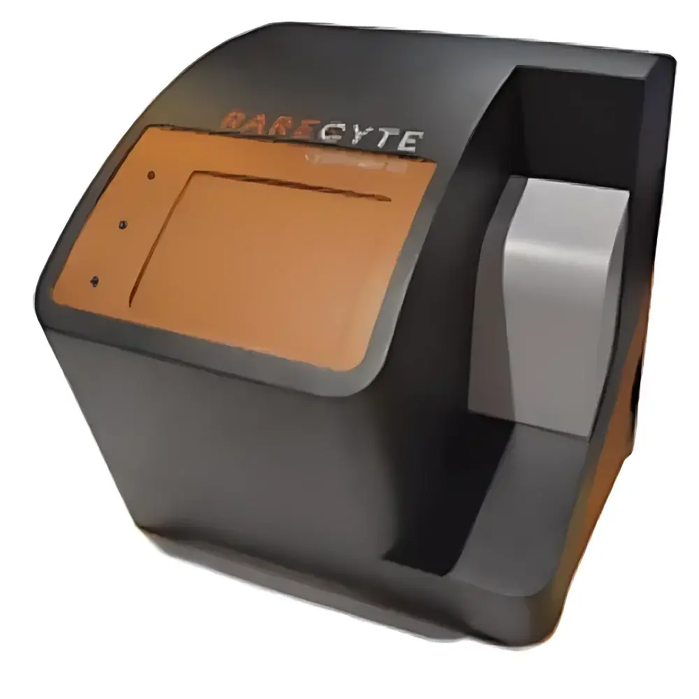
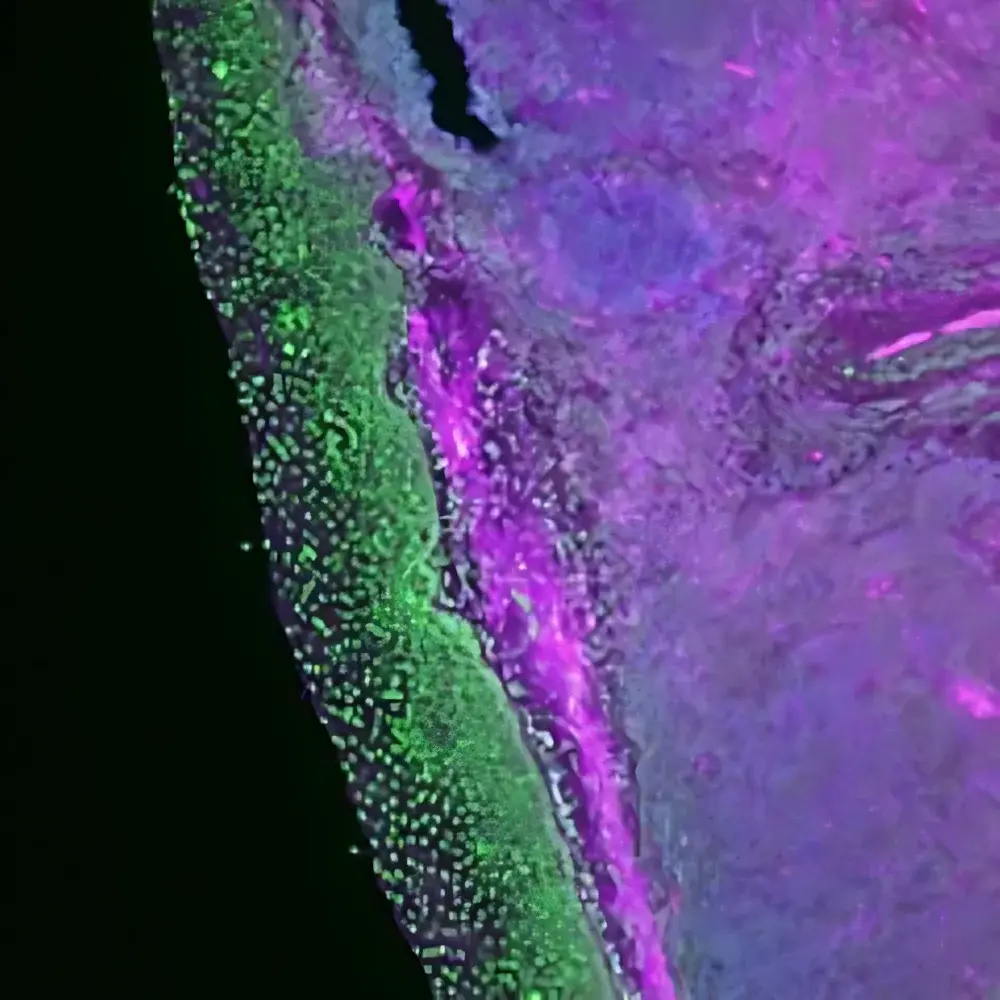
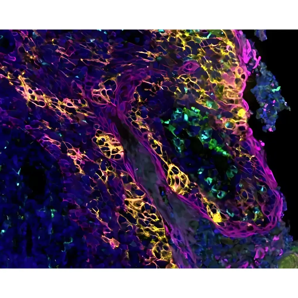
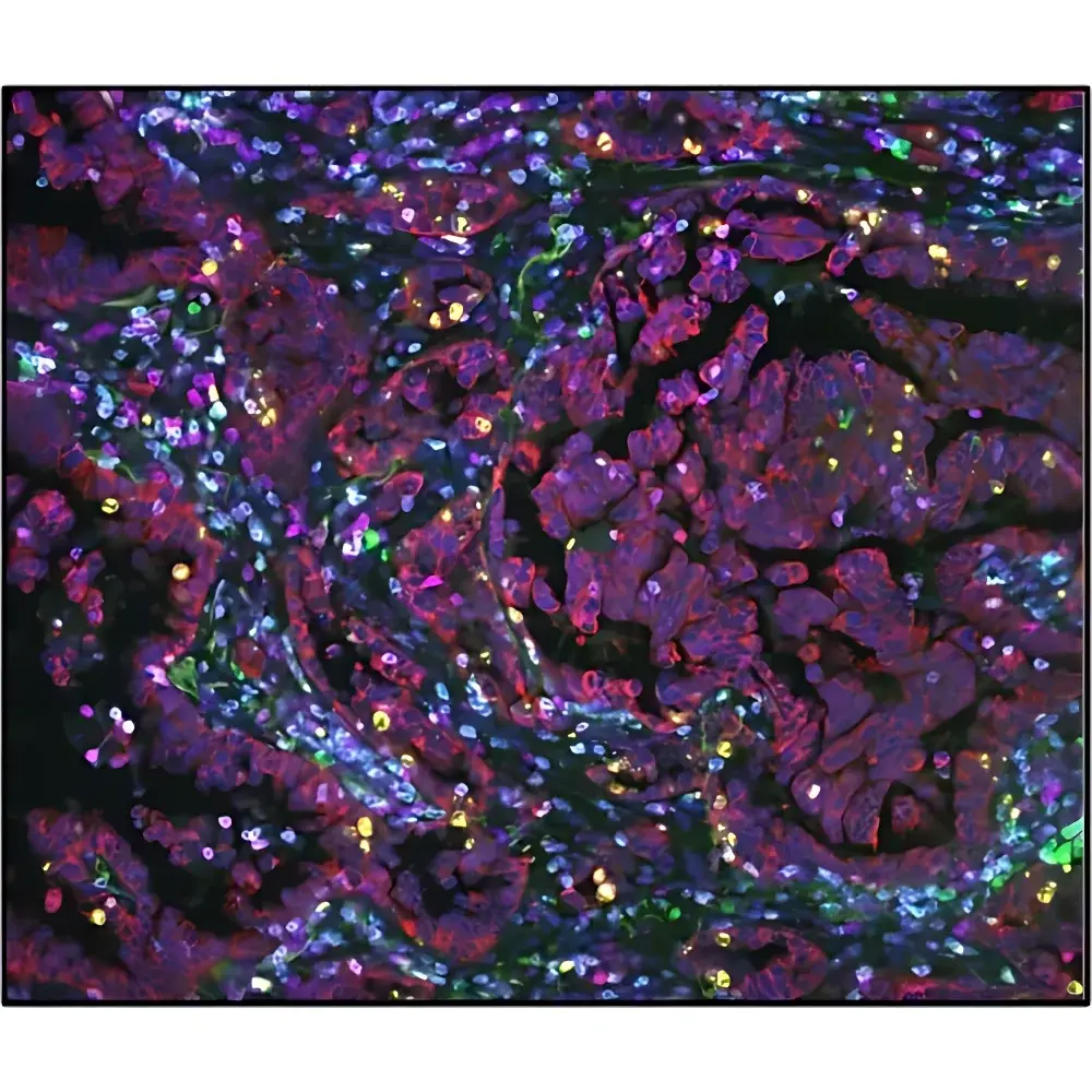
The RareCyte CyteFinder II is an integrated quantitative pathology platform engineered for high-precision, multiplex tissue imaging and image-guided microdissection. Built upon a robust digital pathology architecture, it combines wide-field automated scanning with spectral separation–enabled fluorescence and brightfield imaging to capture spatially resolved molecular and morphological information from formalin-fixed paraffin-embedded (FFPE) and frozen (OCT) tissue sections, cytology preparations, and blood smears. The system operates on a dual-modality acquisition principle: simultaneous multispectral fluorescence imaging (up to 7 channels) and high-fidelity brightfield imaging (including H&E, DAB, and IF), enabling co-registration of protein expression, cellular morphology, and tissue architecture at single-cell resolution. Unlike laser-capture or UV-based microdissection systems, CyteFinder II employs contact-free, non-thermal physical micro-picking—leveraging precision piezoelectric actuation and vacuum-assisted micro-capillary aspiration—to isolate morphologically defined regions of interest (ROIs) without inducing nucleic acid degradation. This preserves native DNA integrity for downstream NGS library preparation and maintains RNA quality for transcriptomic profiling (e.g., RNA-Seq, targeted panel sequencing), forming the technical foundation for Pick-Seq—a validated, image-to-omics workflow for spatially anchored biomarker discovery.
Key Features
- Automated panoramic tissue scanning with sub-micron pixel resolution (≤0.25 µm/pixel at 40× objective), supporting both brightfield and multispectral fluorescence imaging in a single run
- Seven-channel fluorescence capability with hardware-synchronized filter wheel and high-quantum-efficiency sCMOS detection, enabling simultaneous detection of primary antibodies labeled with Alexa Fluor, Cy, and other standard fluorophores
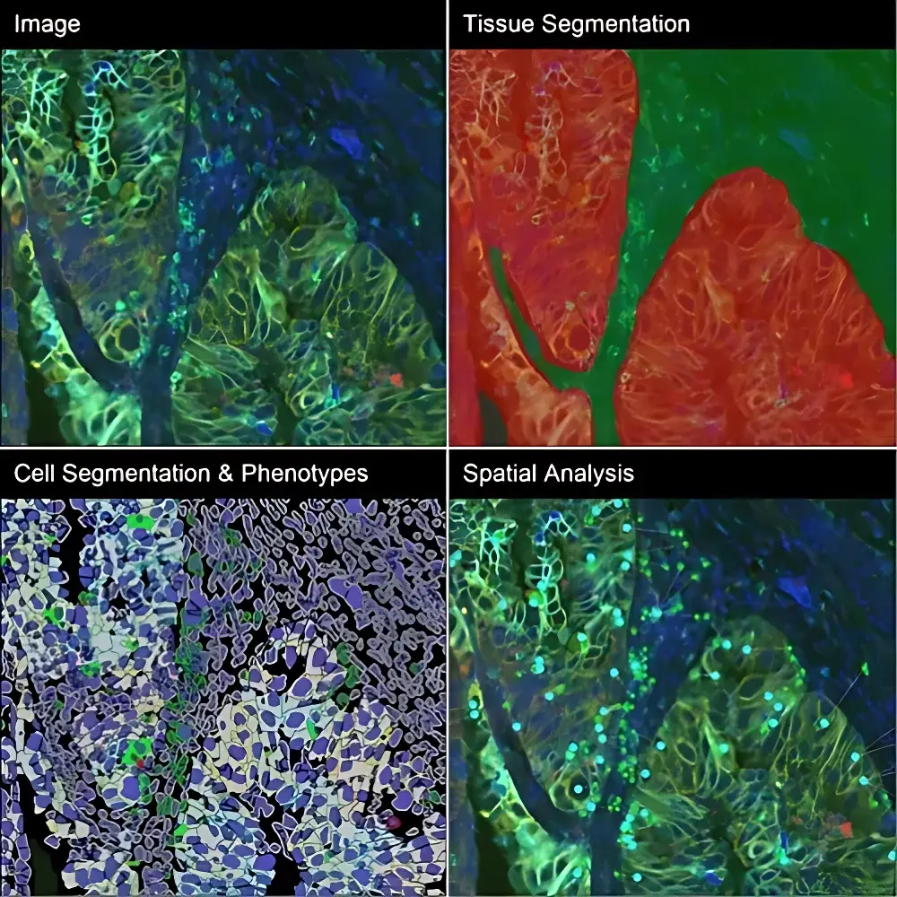
- Integrated CyteHub software suite providing AI-assisted cell segmentation, phenotypic classification (e.g., CD3+/CD8+/PD-1+ T cell subsets), and spatial neighborhood analysis (e.g., tumor-immune interface quantification)
- CytePicker module performing touchless, pressure-controlled microdissection of ROIs as small as 5 µm in diameter—validated for FFPE and OCT sections, cytospins, and membrane slides
- End-to-end traceability: Each microdissected ROI is digitally annotated, georeferenced within the whole-slide image, and assigned a unique barcode for sample tracking through nucleic acid extraction, library prep, and sequencing
- Compliance-ready architecture supporting audit trails, user role permissions, and electronic signatures aligned with GLP and 21 CFR Part 11 requirements for regulated research environments
Sample Compatibility & Compliance
The CyteFinder II accommodates standard histopathology substrates including glass slides (25 × 75 mm), charged and uncharged microscope slides, and specialized membranes used in rare-cell isolation workflows. It supports routine IHC/IF staining protocols (DAB, AEC, fluorescent tyramide signal amplification), hematoxylin and eosin (H&E), and multiplex immunofluorescence panels using Opal™, CODEX®, or custom antibody conjugates. All imaging and microdissection processes are compatible with downstream molecular assays—including whole-exome sequencing (WES), targeted DNA panels, bulk and single-cell RNA-Seq, and methylation analysis—without requiring protocol modification. The platform adheres to ISO 13485 design controls for medical device software components and meets ASTM E2925–21 standards for digital pathology image quality validation.
Software & Data Management
CyteHub serves as the central informatics hub, delivering secure, scalable storage and collaborative annotation of terabyte-scale whole-slide image datasets. Its modular architecture includes: (1) CyteView for real-time image review and ROI markup; (2) CyteAnalyze for unsupervised clustering (PhenoGraph), spatial statistics (Ripley’s K-function), and cell–cell interaction mapping; and (3) CyteExport, which generates MIAME-compliant metadata packages and FASTQ-ready manifests for NGS core facilities. Data encryption (AES-256), SAML 2.0 identity federation, and optional on-premise deployment ensure compliance with HIPAA, GDPR, and institutional data governance policies. Audit logs record all user actions—including image acquisition parameters, ROI selection timestamps, and microdissection event metadata—for full experimental reproducibility.
Applications
The CyteFinder II enables spatially resolved translational research across oncology, immunology, and neuropathology. Key use cases include: characterization of tumor-infiltrating lymphocyte (TIL) functional states (e.g., activation, exhaustion, proliferation) via multiplexed PD-1, TIM-3, LAG-3, Ki67, and FOXP3 co-expression; mapping antigen-presenting cell–T cell synapses in tertiary lymphoid structures; identifying spatially restricted mutational subclones by coupling microdissected ROI genotyping with adjacent section imaging; and validating predictive immune biomarkers (e.g., CD8+/PD-L1+ proximity scores) in clinical trial archival tissues. Its Pick-Seq workflow has been applied in peer-reviewed studies to discover spatially constrained gene signatures associated with checkpoint inhibitor response and resistance.
FAQ
What tissue section thicknesses are supported?
Standard 4–10 µm FFPE and 5–20 µm frozen sections are fully compatible; optimal performance is achieved with 5 µm FFPE and 8 µm OCT sections.
Can CyteFinder II integrate with existing LIMS or ELN systems?
Yes—via RESTful API and HL7/FHIR adapters; pre-built connectors are available for LabVantage, Benchling, and IDBS E-WorkBook.
Is RNA integrity preserved after CytePicker microdissection?
RIN values ≥8.5 have been consistently reported across >200 validated FFPE and frozen samples processed using standard Qiagen RNeasy protocols post-microdissection.
Does the system support batch processing of multiple slides?
Fully automated unattended operation supports up to 12 slides per run with adaptive focus correction and stain-normalized intensity calibration across batches.
Are training and application support services included?
RareCyte provides on-site installation qualification (IQ), operational qualification (OQ), and comprehensive scientist-led training covering imaging optimization, phenotyping pipeline development, and Pick-Seq wet-lab integration.