CAMECA NanoSIMS 50L Secondary Ion Mass Spectrometer
| Brand | Cameca |
|---|---|
| Origin | France |
| Model | NanoSIMS 50L |
| Instrument Type | Magnetic Sector Mass Spectrometer |
| Primary Ion Beam Spot Size | ≥0.5 µm with ion current density ≥100 mA/cm² |
| Mass Range | >238 |
| Mass Resolution (M/ΔM) | >6000 |
Overview
The CAMECA NanoSIMS 50L is a high-performance, magnetic sector-based secondary ion mass spectrometer engineered for nanoscale isotopic and trace elemental analysis with exceptional spatial resolution and analytical fidelity. Operating on the principle of dynamic secondary ion mass spectrometry (SIMS), the instrument utilizes a focused primary ion beam—typically Cs⁺ or O⁻—to sputter material from a solid sample surface; the resulting secondary ions are extracted, energy-filtered, mass-analyzed using a double-focusing magnetic sector analyzer, and detected with high-efficiency multi-collection Faraday cups and electron multipliers. Its coaxial optical design integrates primary beam focusing and secondary ion extraction along a common axis, minimizing aberrations and enabling sub-50 nm lateral resolution in imaging mode. Unlike time-of-flight or quadrupole SIMS systems, the NanoSIMS 50L delivers simultaneous detection of up to seven isotopes at true parallel acquisition—without scanning or pulsing—ensuring high duty cycle, quantitative reproducibility, and intrinsic signal correlation across mass channels.
Key Features
- Sub-50 nm lateral imaging resolution under optimized Cs⁺ or O⁻ primary beam conditions
- Parallel multi-collection capability: simultaneous detection of up to seven isotopes via seven independent detectors (five Faraday cups + two electron multipliers)
- Magnetic sector mass analyzer delivering mass resolution >6000 (M/ΔM) at full transmission, enabling separation of isobaric interferences (e.g., ¹²C¹⁴N⁺ from ²⁶Mg⁺, ³¹P⁺ from ¹⁶O¹⁵N⁺)
- High primary ion current density: ≥100 mA/cm² at beam diameters ≥0.5 µm, supporting high secondary ion yield while preserving spatial fidelity
- Direct-current (DC) acquisition mode—no beam pulsing required—enabling stable, low-noise quantification over extended dwell times
- Integrated charge compensation system for robust analysis of electrically insulating materials (e.g., silicates, polymers, biological tissues)
- Mass range extending from hydrogen (¹H) to transuranic elements (e.g., plutonium isotopes), excluding only noble gases due to ionization inefficiency
Sample Compatibility & Compliance
The NanoSIMS 50L accommodates conductive and non-conductive solid samples—including geological thin sections, polished mineral mounts, freeze-dried biological cells, semiconductor wafers, and ceramic composites—without mandatory metallization. Sample chambers support ultra-high vacuum (UHV) base pressures <1×10⁻⁹ mbar, ensuring minimal surface contamination and stable secondary ion yields. The instrument conforms to ISO/IEC 17025 requirements for testing laboratories when operated within validated analytical protocols. While not FDA-certified per se, its data acquisition architecture supports audit-trail-enabled operation compliant with GLP and GMP documentation standards; raw spectral data files retain full metadata (beam parameters, detector configurations, acquisition timestamps) required for regulatory traceability under 21 CFR Part 11–aligned workflows.
Software & Data Management
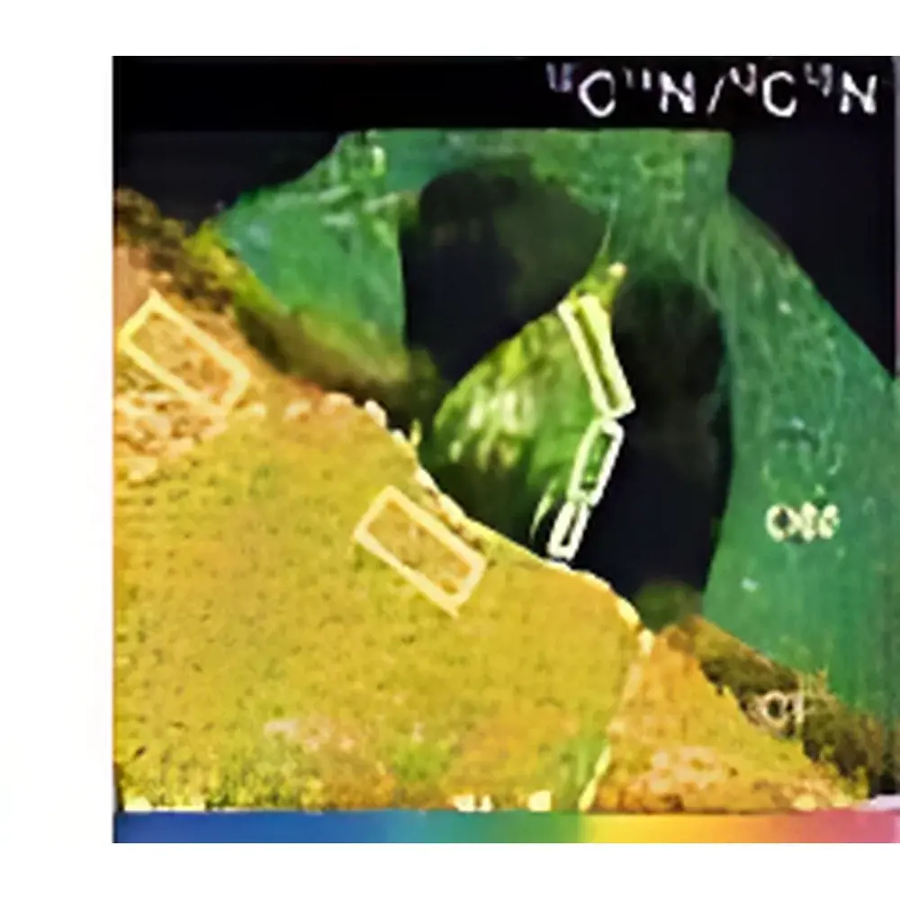
Acquisition and processing are managed through CAMECA’s proprietary L’Image software suite, which provides real-time spectral visualization, interactive mass calibration, drift correction, and pixel-by-pixel isotope ratio mapping. All raw data are stored in vendor-neutral binary formats (.dat/.hdr) compatible with third-party tools such as MATLAB, Python (via open-source libraries like nanosims), and ImageJ/Fiji plugins. Quantitative analysis leverages standard-based or relative sensitivity factor (RSF) methodologies, with built-in tools for dead-time correction, background subtraction, and Poisson noise estimation. Batch processing pipelines support automated ROI definition, ratio image generation (e.g., ¹⁵N/¹⁴N, ¹³C/¹²C), and statistical reporting—including external reproducibility assessment across replicate sessions (typical δ-value precision ≤15‰ for light isotope ratios under controlled conditions).
Applications
- Microbiology & Biogeochemistry: Correlative imaging of phylogenetic identity (via FISH-labeled rRNA) and metabolic activity (via ¹³C-, ¹⁵N-, or ²H-labeled substrates) at single-cell resolution in environmental biofilms or symbiotic consortia.
- Cell Biology: Subcellular tracking of labeled metabolites, lipids, and nucleotides—e.g., monitoring ¹⁵N-amino acid incorporation into organelles or ²H-glucose flux across membrane domains.
- Geosciences & Cosmochemistry: In situ isotopic microanalysis of presolar grains, lunar regolith particles, and zircon inclusion phases—resolving isotopic anomalies (e.g., ²⁶Al/²⁷Al, ⁴⁴Ca/⁴⁰Ca) at <1 µm scale with <0.5% external precision.
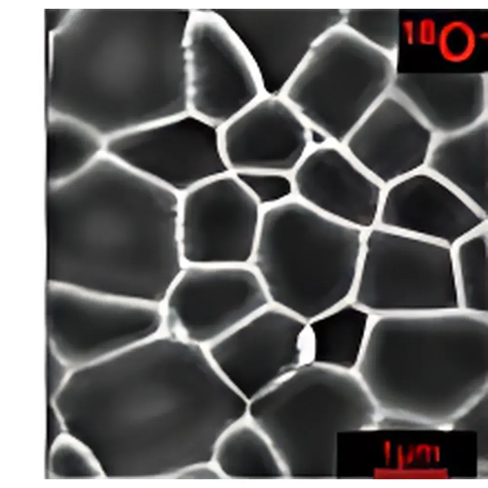
- Materials Science: Dopant profiling in gate oxides, segregation analysis at grain boundaries in high-entropy alloys, and oxidation state mapping in battery cathode materials—leveraging interference-free detection at mass resolution >6000.
FAQ
What is the smallest detectable feature size achievable with NanoSIMS 50L?
The instrument achieves routine lateral resolution of 50 nm in imaging mode under optimal Cs⁺ beam conditions; spot sizes down to 40 nm have been demonstrated in research settings using deconvolution-enhanced acquisition protocols.
Can NanoSIMS 50L analyze insulating samples without conductive coating?
Yes—the integrated electron flood gun and adjustable sample bias enable stable charge neutralization on dielectrics such as quartz, olivine, or frozen-hydrated biological sections.
How many isotopes can be measured simultaneously?
Seven isotopes are acquired in true parallel mode using five Faraday cup detectors and two secondary electron multipliers.
Is mass calibration stable over long-duration acquisitions?
Yes—mass stability is maintained within ±0.1 Da over 24-hour sessions via active temperature-controlled magnet regulation and real-time peak centering algorithms.
What sample preparation methods are recommended for biological specimens?
Cryo-preparation (high-pressure freezing followed by freeze-fracture or cryo-ultramicrotomy) is preferred; resin embedding (e.g., Lowicryl K4M) may be used if cryo-conditions are incompatible with labeling chemistry.
Related Products