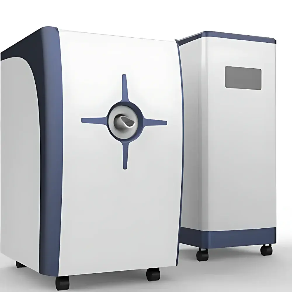
HTNMR HT-AMNMR-50 Small Animal MRI System
| Brand | HTNMR |
|---|---|
| Origin | Shanghai, China |
| Manufacturer Type | OEM Manufacturer |
| Country of Origin | China |
| Model | HT-AMNMR-50 |
| Instrument Type | Benchtop Permanent-Magnet MRI System |
| Animal Model Compatibility | Rat |
| Magnetic Field Strength | 0.3–0.54 T |
| ¹H Resonance Frequency | 18–23 MHz |
| Magnet Bore Diameter | 250 mm |
| Sample Aperture | Φ50 mm |
| Magnetic Field Homogeneity | <8 ppm over 45×45×45 mm³ |
| Image Matrix | up to 256×256×128 |
| Spatial Resolution | down to 0.08 mm (isotropic) |
| Temperature Stability | ±0.06 K/h (after 2-h warm-up) |
| Field Drift | ≤100 Hz/h (Larmor frequency) |
| Maximum Gradient Strength | 70 mT/m (X/Y/Z) |
| Image Linearity | >98% over 50×50×50 mm³ |
Overview
The HTNMR HT-AMNMR-50 is a compact, permanent-magnet-based benchtop MRI system engineered for educational and preclinical research applications. It operates on the fundamental principles of nuclear magnetic resonance—specifically, the detection of radiofrequency signals emitted by hydrogen nuclei (¹H) following excitation in a static magnetic field and controlled gradient encoding. Unlike clinical high-field superconducting MRI systems, the HT-AMNMR-50 utilizes a stable, temperature-regulated permanent magnet assembly with self-shielded gradient coils to minimize eddy current interference—enabling robust implementation of spin-echo, gradient-echo, inversion-recovery, and CPMG-based pulse sequences. Designed for pedagogical rigor and methodological reproducibility, it serves as a platform for teaching MRI physics, validating sequence design, and conducting non-invasive, radiation-free longitudinal imaging studies in rodent models—particularly Sprague-Dawley or Wistar rats within the specified 50 mm sample diameter constraint.
Key Features
- Integrated permanent-magnet system with active temperature stabilization (±0.06 K/h drift after thermal equilibrium), ensuring consistent Larmor frequency stability (≤100 Hz/h drift)
- Self-shielded gradient coil architecture (X/Y/Z; max 70 mT/m) minimizing eddy currents and enabling precise spatial encoding across multi-slice and 3D acquisitions
- Programmable pulse sequence generator supporting custom RF timing, gradient waveform definition, and real-time parameter modulation
- Dual-mode imaging capability: 2D slice-selective acquisition (T₁-weighted, T₂-weighted, proton density) and full 3D volumetric reconstruction (up to 256×256×128 matrix)
- High-fidelity RF probe optimized for ¹H detection at 18–23 MHz, with broadband tuning and matched impedance for SNR optimization
- Comprehensive experimental suite including spin-echo T₂ mapping, inversion-recovery T₁ quantification, gradient-echo contrast evaluation, and IR-based pseudo-color parametric visualization
- Built-in field homogeneity verification module with shimming support across a 45 mm cubic DSV (homogeneity <8 ppm)
Sample Compatibility & Compliance
The HT-AMNMR-50 is validated for in vivo imaging of adult rats (typically 200–400 g) under anesthesia-compatible restraint fixtures. The 50 mm inner diameter sample chamber accommodates standard rat cradles with integrated respiratory gating interfaces. All hardware and software components comply with IEC 61000-6-3 (EMC emission standards) and IEC 61000-6-2 (immunity requirements). While not intended for human use, the system adheres to ISO/IEC 17025-aligned calibration traceability protocols for magnetic field strength, gradient linearity (>98% over 50 mm DSV), and temporal stability. Experimental workflows align with GLP-compliant documentation practices, supporting audit-ready metadata logging—including pulse sequence parameters, acquisition timestamps, and environmental sensor readings (temperature, field drift).
Software & Data Management
The proprietary acquisition and reconstruction software runs on Windows-based host workstations and provides a CLI-accessible API for MATLAB and Python integration. Raw k-space data is stored in vendor-neutral DICOM-RT format with embedded sequence descriptors (TR/TE/TI, flip angle, bandwidth, FOV, matrix size). Reconstruction pipelines include iterative SENSE-based parallel imaging acceleration and GPU-accelerated 3D Fourier inversion. Audit trails record all user-initiated parameter changes with timestamps and operator IDs—meeting foundational requirements for FDA 21 CFR Part 11 compliance when deployed in regulated academic core facilities. Export modules support NIfTI, Analyze, and HDF5 formats for downstream analysis in FSL, AFNI, or SPM environments.
Applications
- Teaching laboratories: Visualization of Bloch equation dynamics, slice selection mechanics, k-space traversal strategies, and relaxation contrast mechanisms
- Biomedical engineering research: Validation of novel pulse sequences, gradient performance benchmarking, and RF coil sensitivity profiling
- Preclinical oncology: Longitudinal tumor volume tracking, necrosis assessment via T₂ mapping, and treatment response monitoring in subcutaneous or orthotopic rat models
- Neuroscience: Ex vivo brain tissue characterization, cerebrovascular flow modeling using phase-contrast gradients, and diffusion-weighted contrast optimization
- Materials science: Non-destructive evaluation of hydrogel swelling kinetics, polymer crosslink density mapping, and porous media fluid distribution analysis
FAQ
What animal models are supported?
The system is optimized for rats (up to 400 g) within a 50 mm cylindrical field-of-view. Mouse imaging is feasible only with reduced FOV and compromised SNR due to hardware aperture constraints.
Is the system compatible with physiological monitoring?
Yes—integrated analog input ports support third-party respiratory and cardiac gating signals via TTL or analog voltage interfaces, enabling motion-compensated acquisitions.
Can users implement custom pulse sequences?
Yes—the open-sequence framework supports user-defined RF/gradient timing tables with microsecond resolution, subject to hardware timing constraints and safety limits.
What level of technical support is provided?
HTNMR offers remote diagnostics, on-site installation commissioning, and annual calibration services performed by certified field engineers trained in MRI safety and magnet quench mitigation protocols.
Does the system meet regulatory requirements for publication-grade data?
All image metadata conforms to DICOM PS3.3 standards, and raw k-space exports retain full phase and magnitude information—satisfying journal requirements for methodological transparency and independent reprocessing.