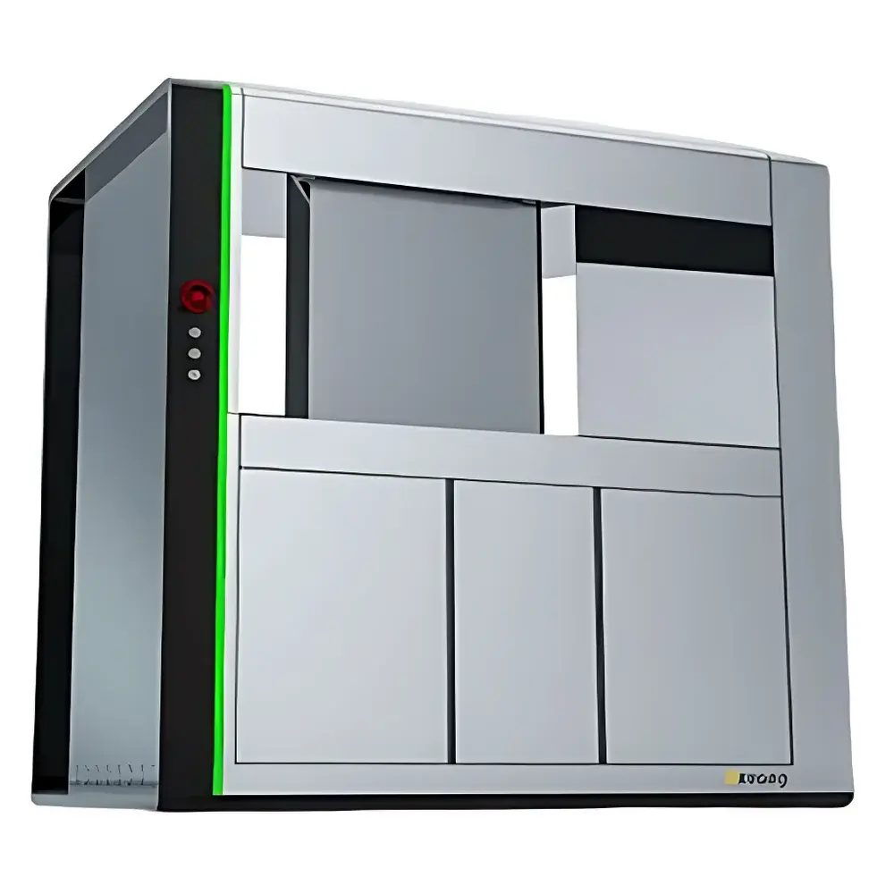
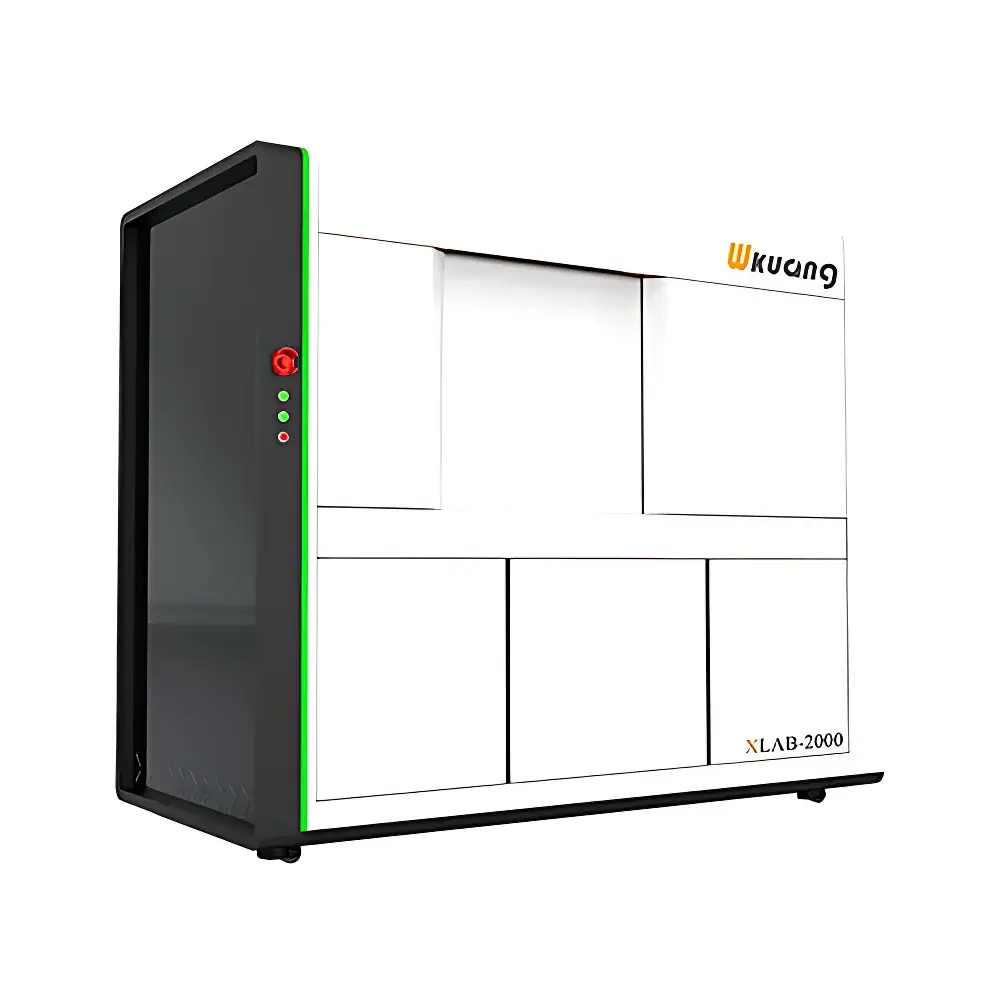
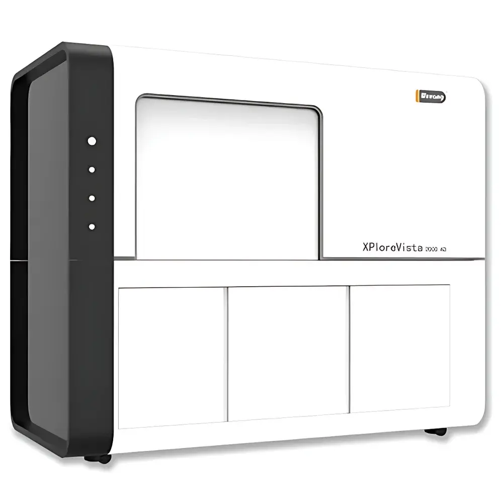
Wkuang XPloreVista 2000 4D Industrial Micro-CT System
| Brand | Wkuang |
|---|---|
| Origin | Jiangsu, China |
| Manufacturer Type | Authorized Distributor |
| Origin Category | Domestic (PRC) |
| Model | XLAB-2000 |
| Quotation | Upon Request |
| Spatial Resolution | ≥0.5 µm |
| Maximum Sample Dimensions | Ø300 mm × H300 mm |
| Dimensional Measurement Accuracy | 200 nm |
| Maximum Sample Weight | 12 kg |
Overview
The Wkuang XPloreVista 2000 4D Industrial Micro-CT System is a high-performance, laboratory-grade X-ray computed tomography platform engineered for non-destructive, quantitative 3D and time-resolved 4D structural analysis of solid materials. Based on cone-beam micro-CT architecture, it employs high-brightness microfocus X-ray source and large-area flat-panel detector to deliver sub-micron spatial resolution (≥0.5 µm) and volumetric reconstruction fidelity suitable for metrology-grade dimensional inspection, pore network characterization, defect detection, and internal architecture validation across advanced manufacturing, materials science, and failure analysis applications. Its dual-contrast imaging capability—integrating absorption-based and phase-contrast modalities—enables reliable visualization of low-Z material interfaces, polymer composites, soft biological tissues embedded in hard matrices, and other structures where conventional attenuation contrast alone yields insufficient contrast-to-noise ratio.
Key Features
- Dual-Contrast Imaging Engine: Combines polychromatic absorption contrast with propagation-based phase-contrast enhancement, leveraging X-ray wavefront interference effects to resolve boundaries between materials with similar linear attenuation coefficients (e.g., carbon fiber/epoxy, alumina/silicon carbide, or hydrated biological phases).
- Automated Multi-Filter Carousel: Integrated 20-position motorized filter wheel enables real-time spectral shaping without interrupting scan acquisition—optimizing beam hardening correction, signal-to-noise ratio, and edge sharpness across heterogeneous samples.
- Modular In-Situ Chamber Architecture: Designed with ≥180 mm internal clearance and standardized flange interfaces (CF100 compatible), the system supports seamless integration of third-party or Wkuang-developed in-situ stages—including high-temperature furnaces (up to 1200 °C), uniaxial/tensile-compression rigs, electrochemical cells, and thermal cycling modules—for true 4D dynamic CT under operational loading conditions.
- High-Precision Mechanical Metrology Subsystem: Equipped with granite base, air-bearing rotation stage, and laser interferometer feedback loop, achieving 200 nm volumetric dimensional measurement accuracy traceable to NIST-traceable artifacts per ISO 15530-3 and VDI/VDE 2634 Part 2 protocols.
- Robust Thermal & Vibration Management: Active temperature stabilization (±0.1 °C) and passive seismic isolation ensure long-duration scans (>24 h) maintain geometric consistency and voxel integrity without drift-induced misregistration.
Sample Compatibility & Compliance
The XPloreVista 2000 accommodates specimens up to Ø300 mm × H300 mm and 12 kg mass—supporting castings, additive-manufactured components, geological cores, battery electrode stacks, ceramic matrix composites, and packaged electronic assemblies. All hardware and software comply with IEC 61000-6-3 (EMC), IEC 61000-6-4 (immunity), and GB/T 18268.1–2010 (industrial measurement equipment safety). Reconstruction workflows adhere to ASTM E1441-22 (Standard Guide for Computed Tomography) and ISO/IEC 17025:2017 requirements for accredited testing laboratories. Optional audit trail logging and user access control align with FDA 21 CFR Part 11 and EU Annex 11 for regulated environments.
Software & Data Management
X-VISON software provides end-to-end processing—from acquisition parameterization to quantification—with support for helical, limited-angle, region-of-interest (ROI), and time-resolved 4D scanning protocols. Embedded algorithms include motion artifact suppression (sub-pixel registration), ring artifact removal (frequency-domain filtering), beam-hardening correction (polynomial-based modeling), and iterative reconstruction (SART + TV regularization). Data output conforms to DICOM-CT, TIFF stack, and HDF5 formats; metadata embedding follows NIEM 4.2 schema. Version-controlled project archives, role-based permissions, and SQLite-backed database indexing enable reproducible analysis and GLP/GMP-compliant documentation.
Applications
- Porosity & inclusion analysis in aerospace turbine blades per ASTM E2987
- In-situ monitoring of crack initiation and propagation during mechanical loading
- Quantitative assessment of binder burnout and sintering densification in MIM parts
- Electrode microstructure evolution in Li-ion pouch cells under cycling stress
- Dimensional verification of dental implants and orthopedic scaffolds against CAD models
- Failure root cause analysis of solder joint voiding, delamination, or intermetallic growth
FAQ
What is the minimum detectable feature size under standard operating conditions?
The system achieves ≥0.5 µm effective spatial resolution at optimal magnification and exposure settings; actual resolvable detail depends on sample composition, geometry, and reconstruction kernel selection.
Does the system support automated batch scanning for QA/QC workflows?
Yes—X-VISON includes scriptable acquisition sequences, barcode-triggered job launching, and pass/fail tolerance mapping against GD&T callouts exported from SolidWorks or CATIA.
Is phase-contrast imaging available as a standard feature or optional add-on?
Phase-contrast mode is fully integrated and enabled by default; no hardware modification or additional licensing is required.
Can raw projection data be exported for third-party reconstruction (e.g., TomoPy, ASTRA Toolbox)?
Yes—unprocessed sinograms and metadata (source-detector geometry, exposure times, filter IDs) are exportable in standard binary+XML format with full calibration parameters.
What is the typical reconstruction time for a 2000×2000×2000 voxel volume?
Using GPU-accelerated FDK reconstruction on an NVIDIA RTX 6000 Ada workstation, full-volume reconstruction completes in ≤8 minutes; iterative methods scale linearly with iteration count and hardware configuration.


