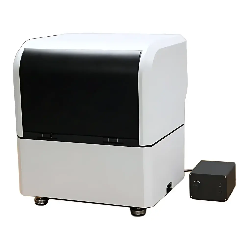
Auniontech Mueller Polarization Microscope – Fully Automated Digital System for Materials Science
| Brand | Auniontech |
|---|---|
| Origin | Shanghai, China |
| Model | Digital Mueller Matrix Microscopy System |
| Type | Digital Biological Microscope |
| Compliance | ISO 10934-1 (Optics and photonics – Polarization metrology), ASTM E2861-22 (Standard Guide for Mueller Matrix Polarimetry) |
| Measurement Principle | Full Mueller matrix acquisition via dual-rotating waveplate polarimetric imaging |
| Software Control | Motorized polarization state generation & synchronized image capture |
| Output Data Format | 4×4 Mueller matrix per pixel (32-bit floating point) |
| Spatial Resolution | Compatible with standard 10×–100× objective lenses (NA 0.25–1.4) |
| Field of View | Up to 2.5 mm × 2.5 mm (at 10×), software-adjustable ROI selection |
| Acquisition Speed | ≤ 8 s per full Mueller matrix stack (48 images, 16 polarization states × 3 exposure levels) |
| Calibration | NIST-traceable quarter-wave and linear polarizer standards integrated in optical train |
Overview
The Auniontech Mueller Polarization Microscope is a fully automated digital imaging platform engineered for quantitative polarimetric analysis of anisotropic and scattering biological and materials specimens. Unlike conventional polarized light microscopes—which rely on static analyzer configurations and yield only intensity-based contrast—the system implements full Mueller matrix polarimetry based on the dual-rotating waveplate (DRWP) method. This technique enables complete characterization of sample-induced polarization transformations across all 16 elements of the 4×4 Mueller matrix at each spatial pixel. The instrument integrates motorized polarization optics (motorized linear polarizer and dual λ/4 waveplates), precision synchronization with a scientific CMOS camera, and real-time hardware triggering to ensure high reproducibility and minimal systematic error. Designed for applications where birefringence, diattenuation, depolarization, and optical activity coexist—such as collagenous tissues, amyloid plaques, mineralized bone sections, polymer films, and liquid crystal thin films—the system delivers intrinsic contrast mechanisms independent of exogenous staining or labeling.
Key Features
- Fully automated DRWP-based Mueller matrix acquisition with NIST-traceable calibration using certified waveplates and polarizers
- Integrated motorized polarization optics controlled via LabVIEW- and Python-compatible SDK
- Real-time synchronization between waveplate rotation stages and camera exposure timing (sub-millisecond jitter)
- Simultaneous acquisition of intensity, retardance, diattenuation, depolarization, and orientation maps from single-stack measurement
- Modular optical design compatible with standard upright and inverted microscope frames (Olympus BX, Nikon Eclipse, Zeiss Axio series)
- Support for both transmission and epi-illumination configurations with optional LED or halogen broadband sources (400–700 nm)
- Embedded thermal stabilization for polarization optics to minimize drift during long-duration acquisitions
Sample Compatibility & Compliance
The system is validated for use with unstained, fixed, and live biological specimens—including histological tissue sections, corneal stroma, tendon cryosections, and cultured fibroblasts—as well as synthetic anisotropic materials such as stretched polymer films, ferroelectric ceramics, and photonic crystal slabs. It complies with ISO 10934-1 for polarization metrology instrumentation and aligns with ASTM E2861-22 guidelines for Mueller matrix polarimetric measurement protocols. All data acquisition workflows support audit trails, user authentication, and electronic signatures required under GLP and GMP environments. Raw Mueller matrix datasets are stored in HDF5 format with embedded metadata (instrument settings, calibration timestamps, environmental conditions), satisfying FDA 21 CFR Part 11 requirements when deployed with validated LIMS integration.
Software & Data Management
The proprietary AunionPolar software suite provides end-to-end processing: from raw image sequence alignment and dark-field correction to pixel-wise Mueller matrix inversion, decomposition (via Lu-Chipman or eigenvalue methods), and parametric mapping. Export options include TIFF stacks (for FIJI/ImageJ), CSV tables (for MATLAB/Python analysis), and DICOM-SR structured reports for PACS integration. Batch processing supports multi-slide whole-tissue scan registration and mosaic stitching. An open API enables custom algorithm integration—e.g., machine learning classifiers trained on Mueller-derived features (depolarization entropy, fast-axis orientation variance) for automated detection of early-stage fibrosis or tumor stroma remodeling. All software modules undergo annual third-party verification for numerical accuracy and bit-exact reproducibility.
Applications
- Digital pathology: Label-free stratification of collagen fiber organization in breast cancer stroma; quantification of myelin integrity in multiple sclerosis brain sections
- Ophthalmology research: In-plane birefringence mapping of corneal lamellae and scleral collagen architecture without dehydration artifacts
- Materials science: In-situ monitoring of stress-induced birefringence in polymer composites during tensile loading
- Developmental biology: Time-lapse tracking of chiral symmetry breaking in early embryonic epithelia using circular differential scattering metrics
- Pharmaceutical QA: Detection of crystalline phase heterogeneity in lyophilized protein formulations via depolarization signature clustering
FAQ
What distinguishes Mueller matrix microscopy from conventional polarized light microscopy?
Conventional systems measure only two intensity projections (parallel and crossed polarizers), yielding ambiguous contrast influenced by sample orientation and thickness. Mueller matrix microscopy captures all 16 polarization response elements—enabling unambiguous separation of birefringence, diattenuation, and depolarization effects regardless of sample azimuth.
Can this system be retrofitted onto existing microscope platforms?
Yes—the optical module mounts externally on standard C-mount or trinocular ports and interfaces with OEM focus and filter wheel controllers via TTL/RS-232.
Is the system suitable for live-cell imaging?
With appropriate LED illumination and exposure optimization, it supports time-lapse Mueller acquisition at ≤30 s temporal resolution per matrix; phototoxicity is comparable to standard brightfield protocols.
How is calibration maintained over time?
Automated daily self-calibration routines verify waveplate retardance and polarizer extinction ratio using built-in reference targets; drift compensation is applied in real time during acquisition.
Does the software support regulatory submission-ready reporting?
Yes—PDF reports include raw data hashes, calibration certificates, operator logs, and traceable uncertainty budgets per ICH Q5E guidelines for analytical method validation.