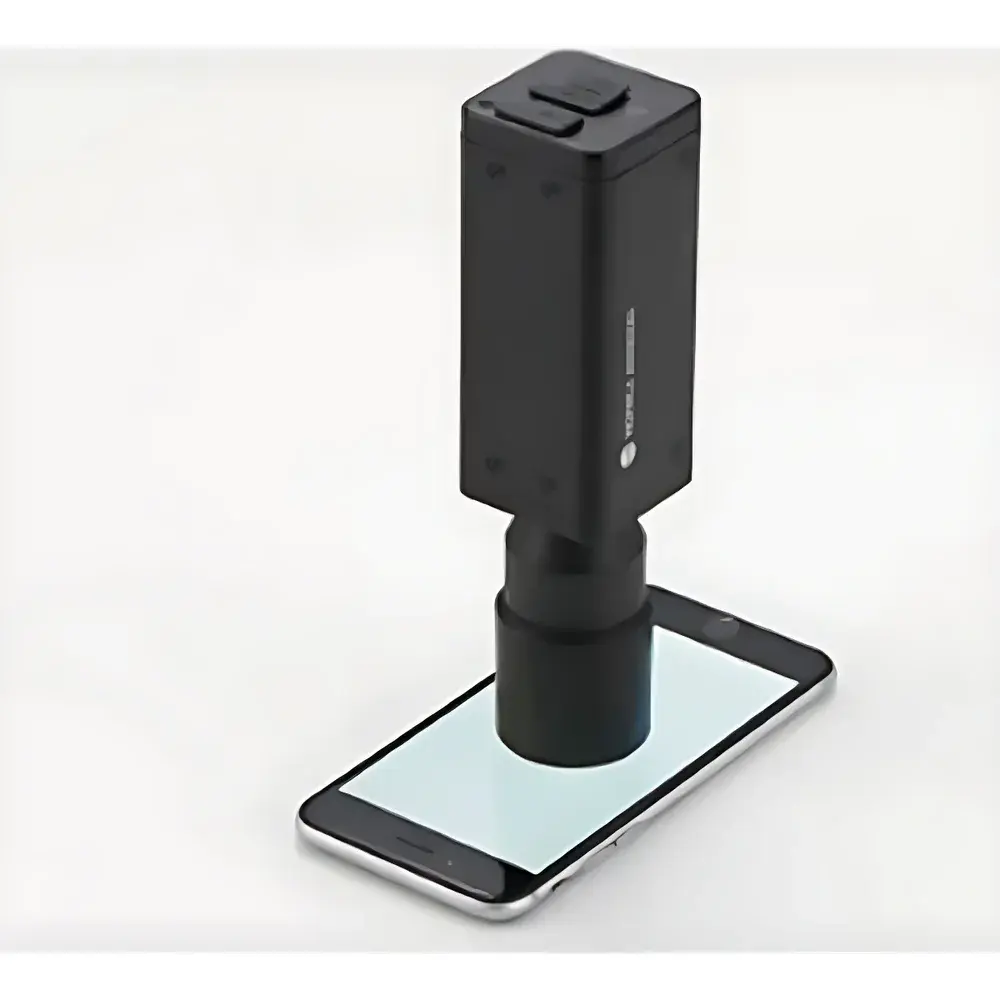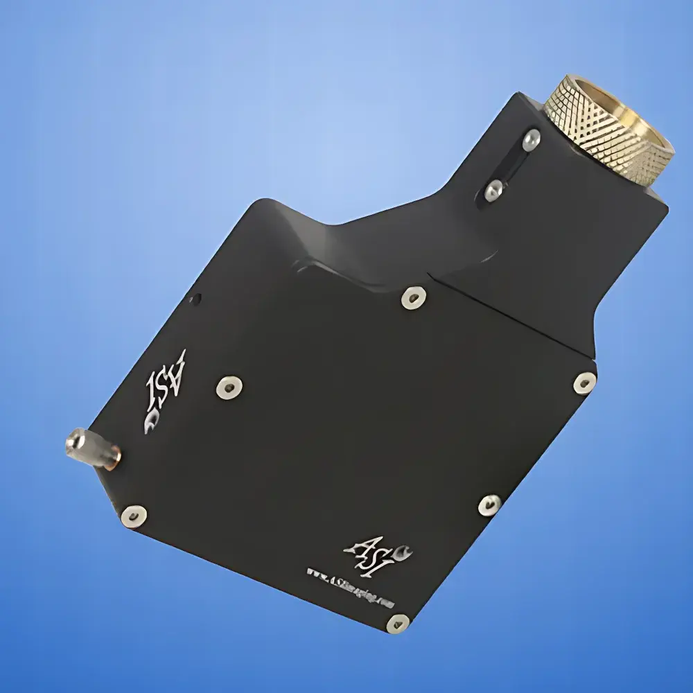
Auniontech CRISP Auto-Focus Module
| Brand | Auniontech |
|---|---|
| Origin | Shanghai, China |
| Model | CRISP Auto-Focus Module |
| Interface | Dual C-Mount Splitter (DCMS) Compatible |
| Optical Principle | Infrared Pattern Projection with Asymmetric Pupil Illumination |
| Feedback Mechanism | Analog Focus Error Signal via Lateral Image Shift Detection |
| Integration | Designed for OEM and research-grade inverted/epifluorescence microscopes |
| Compliance | Compatible with ISO 10110 optical mounting standards and GLP-aligned lab workflows |
| Target Applications | Long-term live-cell imaging, wafer defect inspection, digital pathology slide scanning |
Overview
The Auniontech CRISP Auto-Focus Module is a precision optical distance sensor engineered for real-time, closed-loop maintenance of the objective-to-specimen working distance in high-magnification optical microscopy systems. Unlike conventional contrast- or image-sharpness-based autofocus methods, CRISP operates on a robust physical principle: infrared (850 nm) pattern projection onto the specimen plane through an asymmetrically illuminated half-pupil aperture. This generates a focus-dependent lateral shift in the reflected mask image—directly proportional to defocus error—enabling sub-micron resolution in Z-position feedback without reliance on sample texture or illumination uniformity. The module delivers analog focus error signals at kHz-level bandwidth, making it suitable for dynamic compensation of thermal drift, mechanical creep, and stage-induced axial instability during time-lapse acquisition over hours or days.
Key Features
- Non-invasive infrared illumination (850 nm) minimizes phototoxicity and avoids interference with visible-light fluorescence channels
- Asymmetric pupil illumination architecture ensures stable focus signal generation across low-contrast, transparent, or highly reflective samples—including live cells, silicon wafers, and unstained histological sections
- Analog output interface (±5 V differential) enables direct integration with third-party piezo Z-drivers, motorized objective nosepieces, or custom motion controllers
- Designed for seamless mechanical and optical integration with ASI Dual C-Mount Splitter (DCMS), providing co-registered optical paths for both CRISP sensing and high-resolution digital imaging
- No moving parts in the optical head; solid-state design ensures long-term stability and immunity to vibration-induced misalignment
- Compatible with standard 4x–100x microscope objectives (RMS thread or M27×0.75); supports both air and oil immersion configurations with appropriate DCMS filter sets
Sample Compatibility & Compliance
CRISP maintains focus fidelity across diverse specimen types without algorithmic training or user calibration: adherent and suspension live cells, fixed tissue sections (FFPE and frozen), semiconductor wafers (Si, GaN, SiC), glass substrates, and polymer-based microfluidic devices. Its passive optical sensing mechanism complies with ISO/IEC 17025 requirements for measurement traceability in accredited labs and supports audit-ready documentation when integrated into GLP/GMP-compliant imaging workflows. While CRISP itself does not carry CE or FDA clearance, its modular architecture enables system-level compliance when embedded in Class I or IIa medical device platforms (e.g., digital pathology scanners) under manufacturer-led risk management per ISO 14971.
Software & Data Management
CRISP operates as a hardware-level focus stabilization engine—no proprietary software runtime is required. Analog error signals are acquired via compatible DAQ systems (e.g., National Instruments USB-6211, Thorlabs K-Cube KBD101) and processed using LabVIEW, Python (with PyDAQmx), or MATLAB. Example control scripts—including PID tuning templates and drift-compensation lookup tables—are provided in the developer SDK. For laboratories operating under 21 CFR Part 11 requirements, CRISP’s deterministic analog feedback loop supports electronic signature-capable audit trails when paired with validated data acquisition software and timestamped storage infrastructure.
Applications
- Long-duration live-cell time-lapse imaging (≥24 h) with minimal focus drift (< ±50 nm/hr typical under controlled ambient conditions)
- Automated whole-slide scanning in digital pathology systems requiring consistent Z-positioning across multi-layer tissue sections
- In-line defect inspection of semiconductor wafers during metrology, where thermal expansion induces micron-scale Z-variance between scan lines
- High-resolution confocal and structured illumination microscopy (SIM), where precise Z-registration is critical for optical sectioning fidelity
- Multi-modal correlative microscopy setups combining brightfield, fluorescence, and phase-contrast channels on shared optical pathways
FAQ
How does CRISP differ from laser-based triangulation autofocus systems?
CRISP avoids speckle noise and surface reflectivity dependence by using structured IR projection on the back focal plane—not direct surface interrogation—making it insensitive to sample topography and ideal for biological specimens.
Can CRISP be used with water-dipping or dipping objectives?
Yes—when integrated with a DCMS equipped with appropriate dichroic mirrors and emission filters, CRISP supports upright and inverted configurations with water-immersion objectives up to 60x.
Is real-time Z-scanning possible with CRISP?
CRISP is optimized for focus *maintenance*, not Z-scanning. For dynamic Z-stack acquisition, pair CRISP with a separate high-speed Z-stage and use its error signal as a feedback offset correction layer.
What is the minimum detectable focus shift?
System-level resolution depends on analog signal conditioning and controller bandwidth; typical closed-loop repeatability is ≤100 nm RMS under stable thermal conditions.
Does CRISP require recalibration after objective change?
No—focus error scaling is inherently objective-independent due to pupil-plane referencing; only mechanical alignment of the DCMS is required for new objectives.