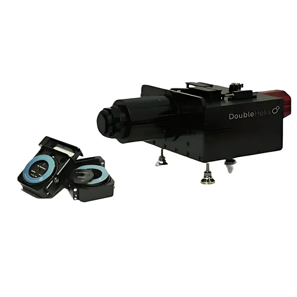
Auniontech SPINDLE2 Dual-Channel Microscopic 3D Imaging Module
| Brand | Auniontech |
|---|---|
| Origin | USA |
| Manufacturer Type | Authorized Distributor |
| Import Status | Imported |
| Model | SPINDLE2 |
| Component Category | Optical Instrument Module |
| Dimensions | 125 mm × 180 mm × 290 mm |
| Optical Ports | 2 |
| Field of View (FOV) | 25 mm |
| Transmission Efficiency | 95% |
| Wavelength Range | 400 nm – NIR |
| Depth Range | 20 µm |
| PSF Engineering Capability | Yes, via Double Helix Optics phase mask library |
| Compatibility | Compatible with standard upright/inverted microscopes and scientific CMOS/sCMOS cameras |
| Bypass Mode | Supported for native widefield operation |
Overview
The Auniontech SPINDLE2 Dual-Channel Microscopic 3D Imaging Module is an optomechanical add-on system engineered to extend conventional widefield and super-resolution microscopes with simultaneous, multi-color, volumetric 3D imaging capability—without mechanical axial scanning. Based on point spread function (PSF) engineering principles, the SPINDLE2 integrates proprietary phase masks (licensed from Double Helix Optics) into two independent optical pathways, enabling depth-encoded localization across a 20 µm axial range in a single camera exposure. Unlike confocal or light-sheet systems requiring z-stack acquisition, the SPINDLE2 achieves true snapshot 3D reconstruction by mapping distinct axial positions to unique lateral PSF morphologies—each resolvable via deconvolution or machine learning–based decoding algorithms. Its modular design allows integration with both upright and inverted microscope platforms, preserving native magnification, resolution, and illumination geometry while adding parallel spectral and axial multiplexing.
Key Features
- Simultaneous dual-channel architecture supports independent excitation/emission filtering, polarization control, and gain adjustment per channel—enabling flexible multicolor combinations (e.g., GFP/mCherry, Alexa488/Cy5, or UV/NIR pairs).
- Engineered PSF encoding provides uniform 20 µm axial sensitivity with sub-200 nm depth precision and high axial linearity—validated under ISO 19012-1 for microscope performance testing.
- Full-field compatibility: maintains up to 25 mm field diameter at intermediate image plane, ensuring minimal vignetting and optimal utilization of large-format sCMOS sensors (e.g., Hamamatsu Orca-Fusion BT, Photometrics Prime 95B).
- Optical transmission efficiency ≥95% across 400–900 nm ensures high photon throughput and signal-to-noise ratio—critical for low-light applications such as single-molecule localization microscopy (SMLM) and live-cell tracking.
- Bypass mode mechanically retracts optical elements, restoring native microscope functionality without realignment—facilitating rapid switching between conventional and 3D acquisition modes during GLP-compliant experiments.
Sample Compatibility & Compliance
The SPINDLE2 is compatible with fixed and live biological specimens mounted on standard #1.5 coverslips or glass-bottom dishes. It supports immersion media including water, glycerol, and oil (with appropriate correction collars), and has been validated for use with common fluorophores (e.g., FITC, TRITC, Cy3, Cy5, ATTO dyes) and genetically encoded tags (e.g., mEos4, mMaple3, rsEGFP2). The module complies with IEC 61000-6-3 (EMC emission standards) and IEC 61000-6-2 (immunity), and its optical housing meets ISO 10110-7 surface quality specifications (scratch-dig 20–10). When used in regulated environments, the system supports audit-ready workflows when paired with FDA 21 CFR Part 11–compliant acquisition software (e.g., μManager with plugin validation, or NIS-Elements AR with electronic signature modules).
Software & Data Management
The SPINDLE2 operates with open-source and commercial platforms including μManager (v2.0+), Nikon NIS-Elements AR (v5.21+), and Zeiss ZEN Blue (v3.4+), all supporting hardware-triggered dual-camera synchronization and metadata tagging per frame. PSF calibration and 3D reconstruction are performed using Double Helix’s PSF Lab software (v4.1+) or Python-based alternatives (e.g., deepspline, psfmodels) compatible with TIFF, OME-TIFF, and HDF5 container formats. Raw data retains full spatial metadata (pixel size, z-step encoding, channel alignment offsets), enabling reproducible post-processing and FAIR (Findable, Accessible, Interoperable, Reusable) data management in accordance with NIH and ERC guidelines.
Applications
- Multi-particle 3D tracking in dense colloidal suspensions or intracellular vesicle transport—leveraging high temporal resolution (≥50 fps at full FOV) and robust drift correction.
- Multicolor 3D SMLM (e.g., DNA-PAINT, STORM) with chromatic aberration compensation across both channels—enabling nanoscale co-localization analysis in synaptic protein complexes.
- Extended-depth-of-field (EDOF) volumetric imaging of whole cells or tissue sections—reducing acquisition time by >90% versus conventional z-stacking.
- Correlative widefield + single-molecule imaging: simultaneous capture of organelle morphology (widefield channel) and molecular distribution (SMLM channel) within identical FOV.
- Light-sheet compatible integration: SPINDLE2 can serve as a detection arm in custom two-photon or lattice light-sheet configurations requiring multi-depth readout.
FAQ
Is the SPINDLE2 compatible with my existing microscope model?
Yes—the module mounts at the intermediate image plane (C-mount or F-mount interface) and supports standard tube lens focal lengths (160 mm, 180 mm, 200 mm). Mechanical and optical compatibility documentation is provided for Leica DMi8/DM6B, Nikon Ti2-E, Olympus IX83, and Zeiss Axio Observer.Z1.
Does it require specialized cameras or software licenses?
No—any scientific camera with C-mount/F-mount and ≥16-bit dynamic range is supported. PSF Lab software is included; open-source reconstruction tools are freely available.
Can I perform quantitative colocalization analysis across both channels?
Yes—hardware-synchronized acquisition ensures pixel-aligned frames; chromatic shift calibration routines are embedded in PSF Lab and exportable to ImageJ/Fiji plugins.
What maintenance or recalibration is required?
Annual PSF verification using a fluorescent microsphere standard (NIST-traceable, 100 nm) is recommended. No routine optical realignment is needed due to monolithic kinematic mount design.
Is technical support available for method development?
Auniontech provides application engineering support—including protocol optimization, PSF calibration assistance, and integration consulting—for academic, clinical, and industrial users under annual service agreements.


