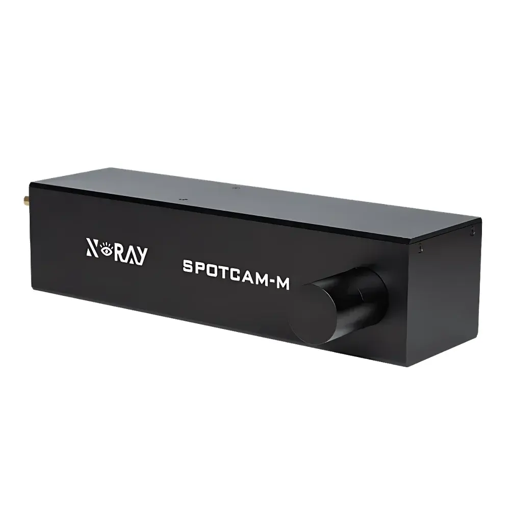
X-rayVision SpotCam-M Lens-Coupled sCMOS X-ray Imaging Camera
| Brand | X-rayVision |
|---|---|
| Model | SpotCam-M |
| Type | Lens-Coupled sCMOS X-ray Detection Camera |
| Detector Technology | Scintillator–sCMOS Optical Coupling |
| Spatial Resolution (Measured) | 0.6 µm (with MH20 lens) |
| Voltage Range Compatibility | 20–70 kV |
| Pixel Array | ≥6200 × 4100 |
| Read Noise | 2 e⁻ (typ.) |
| Dark Current | 0.001 e⁻/pixel/sec @ −17°C (typ.) |
| Cooling | Water-cooled, down to −23°C (@ 20°C ambient) |
| A/D Conversion | 16-bit |
| Exposure Time | 1 ms – 1000 s |
| Interface | USB 3.0 |
| Dimensions | 470 mm × 226 mm × 120 mm |
| Weight | 11.5 kg |
| Lens Options | Manual or Motorized Turret (customizable) |
| Field of View (FoV) | 1.17 mm × 0.78 mm (MH20), 2.34 mm × 1.56 mm (MM10), 5.85 mm × 3.9 mm (ML04) |
| Effective Pixel Size | 0.19 µm / 0.38 µm / 0.95 µm (lens-dependent) |
| Binning Modes | 1×1, 2×2, 3×3, 4×4 |
Overview
The X-rayVision SpotCam-M is a high-performance, lens-coupled sCMOS X-ray imaging camera engineered for quantitative, high-resolution radiography and microtomography in laboratory and industrial X-ray inspection systems. Unlike fiber-optic–coupled or direct-detection sensors, the SpotCam-M employs a precision optical coupling architecture: a high-efficiency scintillator converts incident X-ray photons into visible light, which is then relayed through a selectable high-numerical-aperture (NA) objective lens onto a large-format, low-noise sCMOS sensor. This design preserves spatial fidelity across variable magnifications while enabling optimal signal-to-noise ratio (SNR) at both low-dose and high-frame-rate conditions. The system is optimized for operation with microfocus and nanofocus X-ray sources (20–70 kV), supporting sub-micron resolution imaging in applications demanding metrological traceability and long-term stability—such as semiconductor package inspection, battery electrode microstructure analysis, and additive manufacturing defect characterization.
Key Features
- Sub-micron spatial resolution: Achieves 0.6 µm measured resolution with the MH20 objective lens, validated per ISO 10934-1 methodology using edge-spread function (ESF) analysis.
- Modular optical platform: Supports rapid manual interchange of three calibrated lens options (MH20, MM10, ML04), each pre-aligned and characterized for MTF, distortion, and vignetting—enabling seamless adaptation between high-resolution micro-imaging and wide-field macro-inspection.
- Thermally stabilized sCMOS detection: Integrated water cooling maintains sensor temperature at −23°C (ambient 20°C), suppressing dark current to ≤0.001 e⁻/pixel/sec and ensuring <0.5% pixel response non-uniformity over 1-hour acquisitions.
- Full 16-bit dynamic range with programmable binning: Native 6200 × 4100 pixel array supports on-chip 1×1 to 4×4 binning, balancing resolution, frame rate, and sensitivity without external hardware.
- USB 3.0 interface with deterministic latency: Delivers sustained 120 MB/s throughput and hardware-triggered exposure synchronization (±100 ns jitter), compatible with synchronized multi-camera or source-gating configurations.
- Customization-ready architecture: Optional motorized lens turret integration (with encoder feedback and software-defined positioning) enables automated multi-magnification workflows compliant with ASTM E2737 and ISO/IEC 17025-accredited inspection protocols.
Sample Compatibility & Compliance
The SpotCam-M is designed for use with sealed-tube and open-tube microfocus X-ray sources operating in the 20–70 kV range, including tungsten, molybdenum, and copper anode configurations. Its scintillator selection (Gd₂O₂S:Tb or CsI:Tl, configurable per application) ensures optimal quantum detection efficiency (QDE) across this energy band. The camera meets electromagnetic compatibility (EMC) requirements per EN 61326-1:2013 and carries CE marking for laboratory instrumentation. Mechanical interfaces comply with standard ISO 10360-compliant flange mounts (M42 × 0.75 or C-mount adapters available). For regulated environments—including GMP-compliant battery QA labs or ISO 13485-certified medical device R&D—the system supports audit-trail–enabled acquisition logs and user-access controls when integrated with X-rayVision’s certified acquisition suite (v3.2+), aligned with FDA 21 CFR Part 11 data integrity principles.
Software & Data Management
The SpotCam-M is operated via X-rayVision Acquisition Suite (XAS), a cross-platform (Windows/Linux) application providing real-time image preview, live histogram analysis, flat-field correction, and non-uniformity calibration. Raw images are saved in lossless TIFF or HDF5 format with embedded metadata (exposure time, lens ID, temperature, gain setting, and timestamp). Batch processing pipelines support alignment, drift correction, and SNR-optimized stacking. API access (C/C++, Python bindings) enables integration into custom tomography reconstruction frameworks (e.g., TomoPy, ASTRA) or automated inspection scripts. All calibration files—including lens-specific point-spread function (PSF) maps and gain/dark reference frames—are stored in version-controlled repositories, ensuring reproducibility across instrument deployments and user sessions.
Applications
- High-resolution X-ray microscopy of lithium-ion battery electrodes: Resolving pore networks, particle cracking, and SEI layer morphology at <1 µm scale under operando or ex-situ conditions.
- Semiconductor packaging inspection: Detecting voids, wire bond misalignment, and die attach delamination in QFN, BGA, and fan-out wafer-level packages.
- Materials science research: Quantitative phase-contrast imaging of polymer blends, metal foams, and ceramic composites; correlation with synchrotron-based reference datasets.
- Additive manufacturing quality assurance: In-process and post-build evaluation of internal porosity, unmelted powder, and residual stress-induced microcracks in Ti-6Al-4V and Inconel 718 parts.
- Plasma diagnostics: Time-resolved imaging of X-ray emission from Z-pinch or laser-produced plasmas using gated exposure modes.
FAQ
What scintillator materials are supported, and how do they affect resolution and sensitivity?
The SpotCam-M supports interchangeable scintillators—including Gd₂O₂S:Tb (optimized for 20–50 kV) and CsI:Tl (higher light yield, preferred for 40–70 kV). Thinner scintillators improve spatial resolution but reduce detection efficiency; typical thicknesses range from 10–50 µm. Custom scintillator selection is available upon request.
Is the camera compatible with third-party X-ray sources and motion stages?
Yes. The SpotCam-M provides TTL-compatible trigger I/O and supports GenICam-compliant communication via USB3 Vision protocol extensions. Integration with common motion controllers (PI, Newport, Aerotech) and source modulators (Hamamatsu, Oxford Instruments) is documented in the SDK.
Does the system support real-time tomographic reconstruction?
While the camera itself does not perform reconstruction, its high-throughput USB 3.0 interface and low-latency triggering enable streaming to GPU-accelerated reconstruction engines (e.g., NVIDIA Clara, TomoPhantom) at up to 15 fps for 2k × 2k projections.
How is geometric calibration maintained across lens changes?
Each lens module includes a factory-measured calibration certificate referencing ISO 10360-7. The XAS software automatically loads corresponding distortion coefficients and pixel-scale maps upon lens identification via embedded RFID tags or manual selection.
Can the SpotCam-M be deployed in a cleanroom environment?
The enclosure meets IP52 ingress protection rating. For Class 1000 (ISO 6) cleanrooms, optional stainless-steel housing and filtered air purge ports are available under OEM configuration.
")
")