Dualix GaiaMicro-G Series Microscopic Hyperspectral Imaging System
| Brand | Dualix Spectral Imaging |
|---|---|
| Origin | Beijing, China |
| Model | GaiaMicro-G |
| Spectral Range | 400–1000 nm |
| Spectral Resolution | 3.5 nm |
| Imaging Principle | Push-broom scanning |
| Optical Configuration | Transmission (visible–NIR) and semi-reflective (NIR) |
| Detector Type | CCD/SCMOS/InGaAs |
| Spatial Scanning | Motorized XY precision translation stage |
| Numerical Aperture | F/2.8 (GaiaMicro-G-V10-LU) |
| Slit Size | 30 µm × 9.6 mm |
| Pixel Array | 1392 × 1040 (spatial × spectral) |
| Spectral Channels | 256 (240 effective) |
| Data Depth | 14-bit |
| Interface | USB 2.0 |
Overview
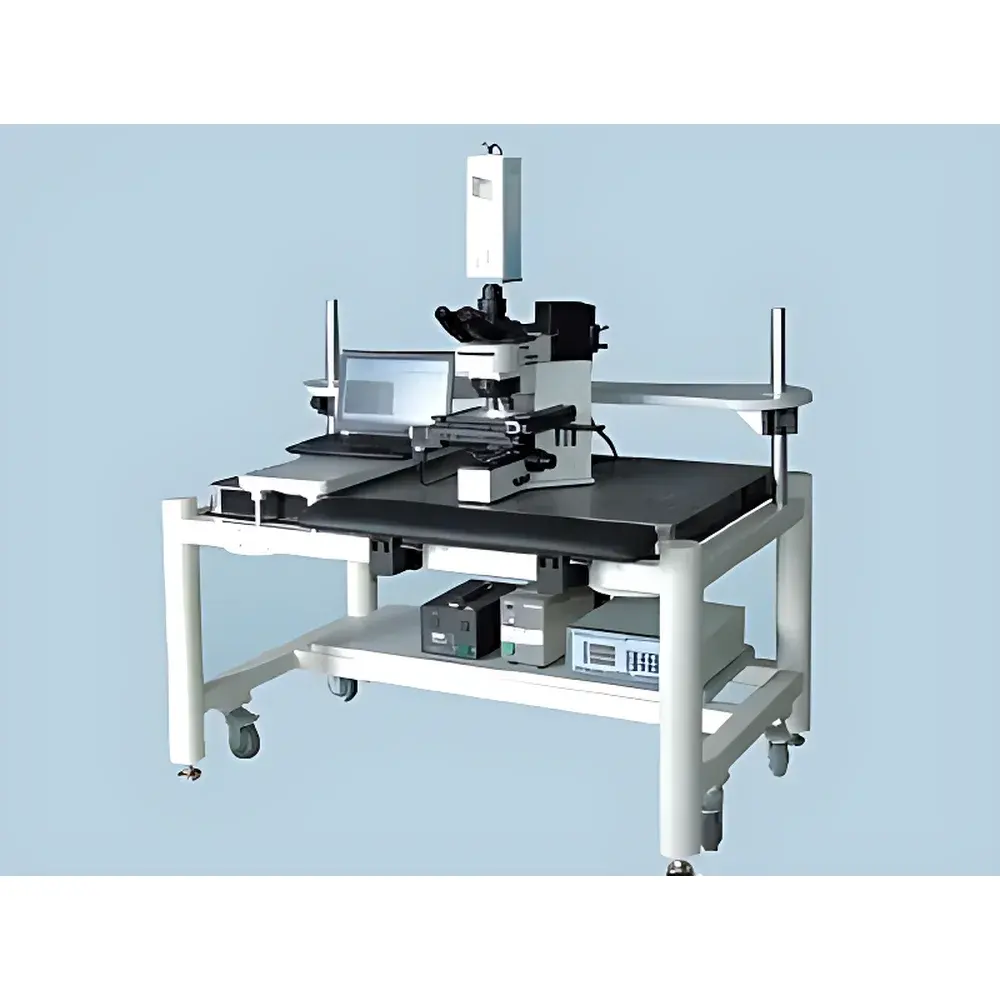
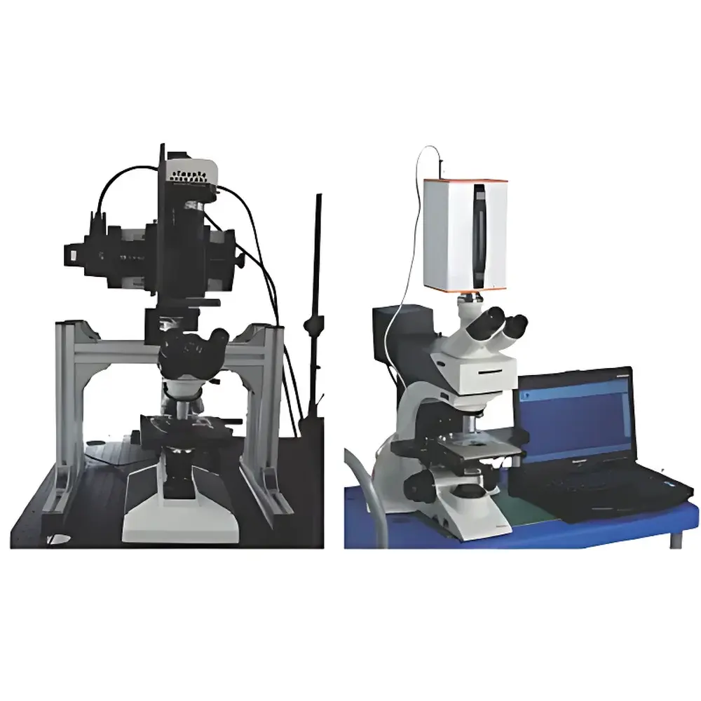
The Dualix GaiaMicro-G Series Microscopic Hyperspectral Imaging System is an engineered integration of push-broom hyperspectral imaging technology with high-precision optical microscopy. Designed for laboratory-based microscale material characterization, it enables spatially resolved spectral acquisition across the visible to near-infrared (VNIR) range (400–1000 nm) with a spectral resolution of 3.5 nm. The system operates on the principle of line-scan spectroscopy: a narrow slit defines the instantaneous field of view, while precise motorized XY translation moves the sample relative to the static spectrometer aperture—capturing a full hyperspectral data cube (x, y, λ) without moving optics. This architecture ensures mechanical stability, high radiometric reproducibility, and minimal optical aberration drift during acquisition. Unlike filter-wheel or tunable-filter systems, the GaiaMicro-G’s push-broom design delivers inherently co-registered spectral bands with consistent point-spread function (PSF) across the entire spectral range—critical for quantitative pixel-wise analysis in materials science, pharmaceuticals, and geoscience applications.
Key Features
- Push-broom acquisition architecture with fixed hyperspectral camera and motorized XY scanning stage—eliminates image registration errors and ensures sub-pixel spatial fidelity.
- Dual optical path configuration: transmission-mode for visible–NIR samples (e.g., thin sections, biological tissues, polymer films) and semi-reflective mode for opaque or reflective specimens (e.g., metallurgical samples, mineral grains, coated surfaces).
- Automated workflow control including auto-focus, auto-exposure, and real-time scan-speed matching based on signal-to-noise ratio (SNR) and illumination uniformity.
- Comprehensive radiometric calibration suite: reflectance calibration using NIST-traceable standards, flat-field correction for illumination non-uniformity, and region-of-interest (ROI)-specific normalization for heterogeneous samples.
- Modular microscope integration: compatible with infinity-corrected finite-conjugate optical systems—including standard achromatic long-working-distance metallurgical objectives (5×, 10×, 20×, 50×), optional 100× reflective objectives, and third-party platforms from Olympus, Zeiss, and Nikon via C-mount or F-mount adapters.
- Precision mechanical stage with ≤100 nm repeatability, programmable velocity profiles, and hardware-triggered synchronization between camera exposure and stage motion.
Sample Compatibility & Compliance
The GaiaMicro-G supports both transmission and reflection modalities, enabling analysis of diverse specimen types: thin-sectioned geological samples, histopathological slides, semiconductor wafers, inked documents, forensic trace evidence, and cultural heritage artifacts. Its optical design accommodates standard microscope slide formats (76 × 26 mm) and custom sample holders up to 50 mm × 50 mm. All calibration protocols adhere to ASTM E275, ISO 13694, and USP <857> guidelines for spectral instrument validation. Data acquisition logs include timestamps, environmental metadata (ambient temperature, humidity), and operator ID—supporting GLP/GMP-compliant documentation requirements. While not FDA 21 CFR Part 11-certified out-of-the-box, the system’s audit trail functionality (via optional software module) enables configuration for regulated environments requiring electronic record integrity and user access controls.
Software & Data Management
Acquisition and preprocessing are managed through Dualix’s proprietary HyperSpectra Studio v4.x—a cross-platform application supporting real-time preview, spectral library matching (ENVI-compatible .sli format), and batch processing workflows. Raw data is saved in HDF5 format with embedded metadata (wavelength vector, slit geometry, objective magnification, NA, exposure time). The software includes built-in tools for spectral unmixing (vertex component analysis), endmember extraction (N-FINDR), and classification (SVM, K-means). Export options include GeoTIFF (with wavelength-indexed bands), CSV (spectral profiles), and MATLAB-compatible .mat files. API support (Python SDK) allows integration into automated QA/QC pipelines and LIMS environments. All calibration files (dark current, flat-field, reflectance reference) are stored with versioned checksums to ensure traceability.
Applications
- Pharmaceutical solid-state analysis: Mapping polymorphic distribution in tablet cross-sections, detecting excipient segregation, and quantifying API concentration gradients at micron-scale resolution.
- Geological thin-section characterization: Discriminating mineral assemblages (e.g., quartz vs. feldspar vs. clay) via diagnostic absorption features near 900 nm and 1400 nm—even when grain sizes fall below conventional microscope resolution limits.
- Materials failure analysis: Identifying oxidation states in corrosion products on metal alloys, mapping carbon black dispersion in rubber composites, and detecting delamination interfaces in multilayer packaging films.
- Biomedical research: Label-free identification of collagen cross-linking in connective tissue, spectral differentiation of necrotic vs. viable tumor regions in frozen sections, and pigment distribution analysis in retinal models.
- Forensic document examination: Differentiating ink chemistries across overlapping handwritten entries, revealing erased or altered text through subtle spectral shifts in dye degradation products.
FAQ
What is the minimum resolvable feature size under 50× magnification?
At 50× with F/2.8 NA and 400–1000 nm spectral coverage, theoretical diffraction-limited lateral resolution is ~0.35 µm (at 400 nm); practical system resolution depends on detector pixel sampling and objective MTF—typically 0.5–0.8 µm in calibrated operation.
Can the system be upgraded to cover SWIR (1000–2500 nm)?
Yes—by replacing the VNIR camera module with an InGaAs detector and integrating a 900–1700 nm optimized GaiaMicro-G-N17E variant, including matched reflective objectives and broadband halogen illumination.
Is remote operation supported for cleanroom or hazardous environments?
All motorized components and camera interfaces support Ethernet-based control (GigE Vision compliant on select models); optional fiber-optic shutter and LED illumination modules enable fully isolated operation.
How is spectral calibration verified over time?
A built-in mercury-argon emission lamp module (optional) provides periodic wavelength reference checks; factory calibration certificates include uncertainty budgets per ISO/IEC 17025 requirements.
Does the software support batch processing of multi-sample datasets?
Yes—HyperSpectra Studio includes scriptable batch mode with customizable ROI templates, spectral index calculations (e.g., NDVI, PRI), and export to relational database schemas via ODBC connectors.