Auniontech Model 4200M Microscopic Staring Hyperspectral Imaging System
| Brand | Auniontech |
|---|---|
| Origin | Imported |
| Model | 4200M |
| Spectral Range | 400–1000 nm |
| Spectral Channels | 300–600 |
| Spectral Resolution | 4 nm |
| Spatial Resolution | 2.3 MP (1920 × 1200) |
| Bit Depth | 8 or 16 bit |
| Interface | USB 3.0 |
| Operating Temperature | 20 °C ± 5 °C |
| Relative Humidity | ≤65% non-condensing |
| Power Supply | 18 VDC (optical head only) |
| Dimensions | 230 × 120 × 200 mm |
| Weight | 1.4 kg |
| Imaging Mode | Snapshot (staring), 3D spectral data cube acquisition |
| Platform Compatibility | Benchtop microscope integration |
Overview
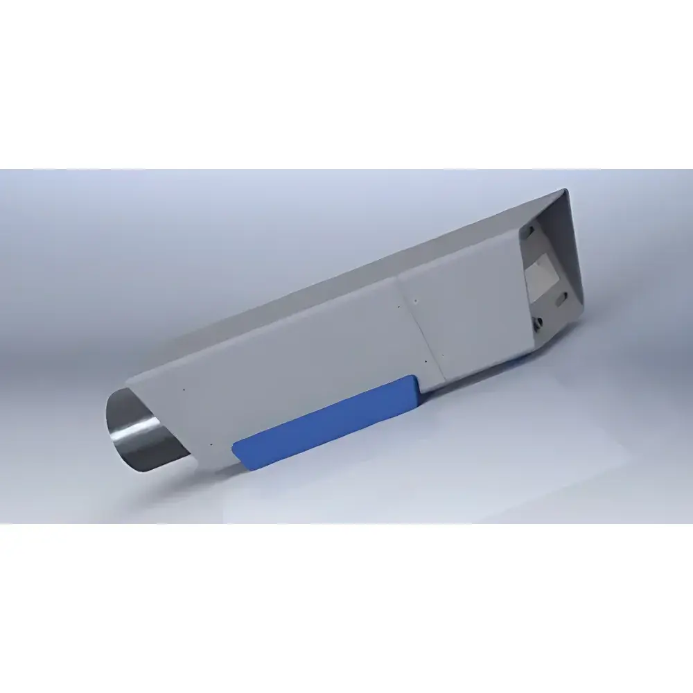
The Auniontech Model 4200M Microscopic Staring Hyperspectral Imaging System is a modular, snapshot-capable hyperspectral add-on module engineered for integration with standard upright and inverted optical microscopes. Unlike scanning-based or filter-wheel-driven multispectral systems, the 4200M employs a staring (area-scan) architecture based on a high-throughput, tunable spectral filtering mechanism—enabling full-frame acquisition of spatially registered spectral data cubes without mechanical scanning or time-multiplexed exposure. This design eliminates motion artifacts, preserves native spatial resolution, and supports real-time spectral analysis at the subcellular and nanomaterial scale. The system operates across the visible to near-infrared (VIS-NIR) spectrum (400–1000 nm), delivering up to 600 discrete spectral bands with a consistent 4 nm full-width-at-half-maximum (FWHM) resolution. Its compact form factor (230 × 120 × 200 mm, 1.4 kg) and low-power 18 VDC requirement allow seamless integration into regulated laboratory environments—including GLP-compliant histopathology labs and ISO 17025-accredited materials testing facilities.
Key Features
- Snapshot Staring Architecture: Captures full 2.3 MP (1920 × 1200) spatial frames with simultaneous spectral sampling—no line scanning, no stage movement, no spectral registration drift.
- Programmable Spectral Subset Acquisition: Users define custom wavelength ranges and channel spacing via software API; eliminates mandatory full-cube acquisition—reducing file size by up to 70% and accelerating throughput in high-volume screening (e.g., tissue microarray analysis).
- Preserved Spatial Fidelity: Avoids the spatial resolution degradation inherent in filter-array-based multispectral cameras (e.g., Bayer- or mosaic-CFA sensors); maintains diffraction-limited performance when coupled with high-NA microscope objectives.
- Flexible Integration Pathway: Designed as a C-mount or infinity-corrected tube lens interface module; compatible with major OEM microscope platforms (Olympus, Nikon, Zeiss, Leica) and third-party motorized stages.
- Low-Light Optimized Optics: High transmission efficiency (>65% avg. across 400–1000 nm) and low read noise (<12 e⁻ RMS) support fluorescence, reflectance, and transmittance modalities—even under LED or halogen illumination.
- Regulatory-Ready Data Handling: Supports timestamped metadata embedding (EXIF + custom XML), hardware-triggered acquisition sync, and optional audit-trail logging compliant with FDA 21 CFR Part 11 requirements when deployed with validated software workflows.
Sample Compatibility & Compliance
The 4200M accommodates diverse sample types—from fixed and live biological specimens (e.g., stained tissue sections, cultured cells, biofilms) to solid-state materials (polymers, thin-film coatings, semiconductor wafers), particulates (microplastics, soil aggregates), and heritage artifacts (pigment layers, ink formulations). It operates under ambient lab conditions (20 °C ± 5 °C, ≤65% RH non-condensing) and requires no vacuum or cryogenic support. All optical components comply with RoHS 2011/65/EU and REACH SVHC directives. When integrated into microscopy workflows, the system supports ASTM E2821-21 (standard guide for hyperspectral imaging in forensic document examination) and ISO 18566-2:2020 (spectral imaging for cultural heritage documentation). For pharmaceutical applications, spectral calibration traceability aligns with USP Photometric Absorbance and ICH Q5C stability guidelines.
Software & Data Management
The system ships with Auniontech’s Hyperspectral Studio v4.x—a cross-platform application (Windows/Linux) supporting real-time preview, spectral library matching (NIST SRD 143, USGS Spectral Library), endmember extraction (VCA, N-FINDR), and quantitative unmixing (linear constrained least squares). Raw data is saved in vendor-neutral ENVI-compatible .hdr/.dat format with embedded geospatial and acquisition metadata. An open-source Python SDK (hyperspy-compatible) enables integration with scikit-learn, TensorFlow, and PyTorch for supervised classification (e.g., tumor vs. stroma segmentation) or autoencoder-based denoising. All processing pipelines support batch-mode execution and can be exported as reproducible Jupyter notebooks—facilitating method transfer between QC labs and satisfying ALCOA+ (Attributable, Legible, Contemporaneous, Original, Accurate, Complete, Consistent, Enduring, Available) data integrity principles.
Applications
- Life Sciences: Multiplexed fluorescence imaging using spectrally overlapping dyes (e.g., Alexa Fluor 488/532/568), microenvironment mapping via Stokes shift heterogeneity, label-free cellular phenotyping based on intrinsic absorption signatures.
- Clinical Pathology: Digital spectral histology for margin assessment in frozen-section analysis; detection of amyloid deposits, collagen cross-linking, or lipid accumulation without staining.
- Materials Characterization: Polymer blend phase segregation analysis, corrosion product identification on metallic substrates, graphene layer counting via interference contrast modeling.
- Forensics: Non-destructive ink differentiation on questioned documents, latent fingerprint enhancement via spectral contrast, accelerant residue discrimination in arson debris.
- Environmental Monitoring: Microplastic polymer typing in wastewater filters, chlorophyll-a/carotenoid ratio quantification in algal blooms, heavy metal speciation mapping in soil thin sections.
- Cultural Heritage: Pigment stratigraphy reconstruction in painted wood panels, binder identification in historical manuscripts, varnish aging assessment via carbonyl band tracking.
FAQ
Is the 4200M compatible with confocal or super-resolution microscopes?
Yes—when installed at the intermediate image plane (e.g., side port or camera port), it maintains compatibility with laser-scanning confocal systems and structured illumination platforms, provided optical path length and magnification are preserved per manufacturer specifications.
Does the system require external calibration sources?
Factory-calibrated radiometric and spectral response data is embedded in firmware; however, optional NIST-traceable tungsten-halogen and mercury-argon lamp kits are available for routine verification per ISO/IEC 17025 internal quality control protocols.
Can spectral data be exported for third-party analysis (e.g., MATLAB, R)?
Yes—raw cube data exports in IEEE 754 floating-point .raw format with header files readable by MATLAB’s hyperspectral toolbox, R’s hyperSpec package, and ENVI Classic/IDL.
What microscope objective magnifications are supported?
The system supports all standard finite-conjugate and infinity-corrected objectives from 2× to 100×; optimal performance is achieved with NA ≥ 0.4 and working distance ≥ 1 mm to avoid vignetting.
Is airborne deployment validated?
While the optical head meets MIL-STD-810G shock/vibration thresholds, airborne integration requires additional environmental enclosure, GPS-synchronized triggering, and custom mounting brackets—available under engineering services agreement.