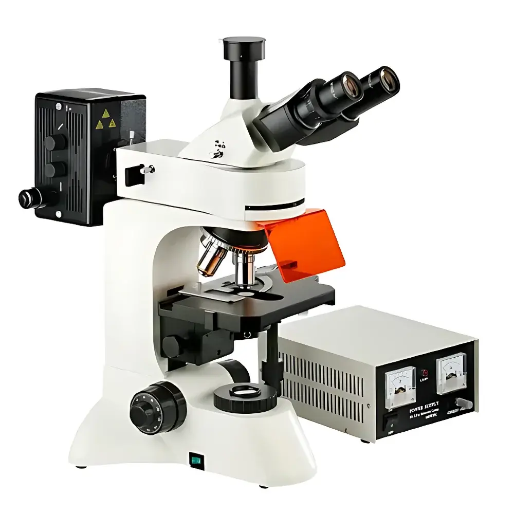
Betop Scientific L3201 Upright Fluorescence Microscope
| Brand | Betop Scientific |
|---|---|
| Origin | Guangdong, China |
| Manufacturer Type | Direct Manufacturer |
| Product Category | Domestic |
| Model | L3201 |
| Instrument Type | Upright Fluorescence Microscope |
| Excitation Source | 100 W High-Pressure Mercury Lamp |
| Medical Device Classification | Non-Medical Device |
| Microscope Class | Conventional Epifluorescence Microscope |
| Eyepieces | Wide-Field WF10X (Φ22 mm) |
| Objectives | Infinity-Corrected Achromatic Plan Objectives |
| Fluorescence Filter Sets | UV, Violet, Blue, and Green Excitation Filter Cubes |
| Illumination System | Dual Illumination (Epifluorescence + Transmitted Light) |
| Control Mode | Manual |
| Focusing Mechanism | Coaxial Coarse/Fine Focus with 2 µm Fine Adjustment Graduation, Locking & Limit Stop |
| Condenser | Abbe Condenser, N.A. 1.25, Vertical Adjustment |
| Observation Head | Trinocular, 30° Inclination |
| Stage | Double-Layer Mechanical Stage, 210 mm × 140 mm, Travel Range: 75 mm × 50 mm |
| Fluorescence Filters | Blue Excitation Filter + Ground Glass Diffuser |
| Optional Accessories | Reticle Eyepieces, 0.4X/0.5X/1X C-Mount Adapters, USB CMOS Cameras (1.3–9 MP), Canon EF/Nikon F Mount Adapters |
Overview
The Betop Scientific L3201 Upright Fluorescence Microscope is engineered for reliable epifluorescence imaging and transmitted brightfield observation in routine life science laboratories. Built upon an infinity-corrected optical architecture, the system delivers high-fidelity image formation with minimal chromatic aberration and optimized flat-field performance across the entire field of view. Its modular design supports standardized fluorescence excitation pathways—UV, violet, blue, and green—enabling multi-channel detection of common fluorophores including DAPI, FITC, TRITC, and Alexa Fluor dyes. The microscope employs a 100 W high-pressure mercury lamp as the primary excitation source, delivering stable spectral output across key UV–visible bands required for conventional fixed- and live-cell fluorescence applications. Designed for ergonomic operation, the L3201 features a low-position coaxial focus control, inward-facing quintuple objective turret with ball-bearing positioning, and a 30° inclined trinocular head compatible with photomicrography and digital documentation systems.
Key Features
- Infinity-corrected plan achromatic objectives (4×, 10×, 40×, 100× oil) ensure uniform resolution and minimal field curvature, critical for quantitative fluorescence intensity assessment.
- Wide-field 10× eyepieces (Φ22 mm field number) provide extended visual coverage and reduced eye strain during prolonged observation sessions.
- Dual illumination capability: epifluorescence path with four standard filter cubes and Köhler-optimized transmitted halogen illumination (6 V / 30 W) with adjustable brightness and centerable field diaphragm.
- Abbe condenser (N.A. 1.25) with vertical adjustment enables precise aperture control for optimal contrast in both brightfield and phase contrast modes (when equipped with optional phase components).
- Double-layer mechanical stage (210 mm × 140 mm) offers smooth, backlash-free movement over 75 mm × 50 mm range—designed for reproducible specimen repositioning and time-lapse coordinate tracking.
- Integrated safety features include lamp power regulation, heat-absorbing filters in the fluorescence path, and mechanical limit stops on fine-focus mechanism to prevent objective collision.
Sample Compatibility & Compliance
The L3201 accommodates standard glass slides (26 mm × 76 mm), Petri dishes (up to 100 mm diameter), and multi-well plates (e.g., 24- or 96-well formats) when used with appropriate stage adapters. It supports both fixed and live specimens—including cultured mammalian cells, yeast, bacteria, tissue sections, and zebrafish embryos—under ambient or controlled environmental conditions. While not certified as a medical device under FDA 21 CFR Part 809 or ISO 13485, the instrument meets general laboratory equipment requirements per IEC 61010-1 for electrical safety and electromagnetic compatibility. Its optical configuration aligns with ASTM E2877-22 guidelines for fluorescence microscope verification and supports GLP-compliant documentation workflows when paired with validated digital imaging software.
Software & Data Management
The L3201 is fully compatible with third-party scientific imaging platforms via standard C-mount interfaces (0.4×, 0.5×, 1× reduction options) and USB 2.0/3.0 outputs supporting resolutions up to 9 megapixels. Supported camera protocols include UVC-compliant drivers for Windows/Linux/macOS environments. Image acquisition, annotation, measurement (length, area, intensity profiling), and multi-channel overlay functions are enabled through open-architecture software such as ImageJ/Fiji, CellSens Standard (Olympus), or NIS-Elements (Nikon). Audit trail functionality—including timestamped metadata capture, user login logging, and raw data export in TIFF/OME-TIFF formats—can be implemented in accordance with internal SOPs for quality-controlled research environments.
Applications
- Subcellular localization studies using fluorescent protein tags (e.g., GFP, mCherry) in transfected cell lines.
- Immunofluorescence detection of antigen distribution in formalin-fixed paraffin-embedded (FFPE) or frozen tissue sections.
- Microbial identification and viability assessment via nucleic acid stains (e.g., SYTO 9/PI) or membrane potential dyes.
- Autofluorescence screening of plant tissues, metabolic intermediates, or endogenous fluorophores in clinical cytology samples.
- Time-lapse imaging of dynamic processes such as mitosis, organelle trafficking, or calcium signaling—when combined with environmental chamber accessories.
FAQ
Is the L3201 suitable for quantitative fluorescence intensity measurements?
Yes—its infinity-corrected optics, calibrated excitation filter sets, and stable mercury lamp output support semi-quantitative comparisons across samples when acquisition parameters (exposure time, gain, binning) are rigorously controlled.
Can oil immersion objectives be used with the standard condenser?
Yes—the Abbe condenser (N.A. 1.25) is matched to the 100× oil objective (N.A. 1.25) for optimal resolution in both brightfield and fluorescence modes.
Does the system comply with FDA 21 CFR Part 11 for electronic records?
The microscope itself does not embed electronic record functionality; however, when integrated with compliant imaging software and networked storage systems, it supports Part 11-aligned audit trails and electronic signatures.
What camera interfaces are supported for digital imaging?
C-mount adapters (0.4×, 0.5×, 1×) and USB-based CMOS cameras (1.3–9 MP) with UVC drivers are natively supported; DSLR adapters for Canon EF and Nikon F mounts are available as optional accessories.
Is service and technical support available outside China?
Betop Scientific provides international distributor networks with local calibration, maintenance, and application support services—contact regional partners for warranty and repair coordination.
Related Products