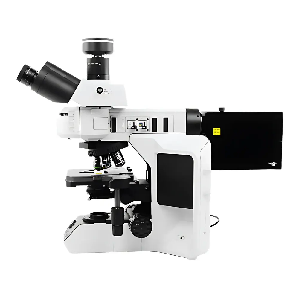
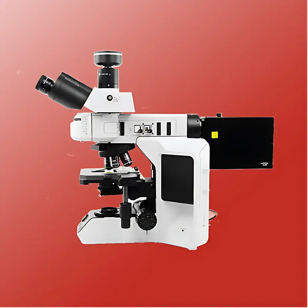
Betop Scientific TX53F Research-Grade Upright Fluorescence Microscope
| Brand | Betop Scientific |
|---|---|
| Origin | Guangdong, China |
| Manufacturer Type | Direct Manufacturer |
| Instrument Type | Upright Fluorescence Microscope |
| Model | TX53F |
| Excitation Source | High-Power LED |
| Microscope Category | Research-Grade Fluorescence Microscope |
| Eyepieces | Widefield 10×, Field Number (FN) = 23 |
| Objectives | 4×/0.13, 10×/0.30, 20×/0.50, 40×/0.75, 100×/1.3 (Oil Immersion) |
| Fluorescence Illumination | High-power solid-state LED source with pre-installed four-wavelength excitation (340–750 nm range) |
| Transmitted Light Source | Full-spectrum high-CRI LED illuminator (60,000 h lifetime, 4500 K CCT) |
| Focus Mechanism | Manual coaxial coarse/fine focus (coarse travel: 17.6 mm/rev |
| fine resolution | 1 µm per division |
| total vertical travel | 27 mm) |
| Condenser | Abbe condenser, NA = 1.1, optimized for 4×–100× objectives |
| Observation Head | Trinocular widefield head with dual beam-splitting ratio (0/100 or 50/50), FN = 23 |
| DIC Compatibility | Integrated DIC slot and polarizer/analyzer mounting capability |
| Stage | Manual mechanical stage with nano-ceramic anti-wear coating, right-hand XY coaxial control, adjustable damping ring |
| Fluorescence Filter Sets | (1) UV broadband excitation block |
Overview
The Betop Scientific TX53F is a research-grade upright fluorescence microscope engineered for high-fidelity cellular and subcellular imaging in life science laboratories. Built upon an advanced ECGO infinity-corrected optical system with bidirectional chromatic aberration correction, the TX53F delivers exceptional image contrast, color fidelity, and spatial resolution across its full magnification range (40×–1000×). Its optical architecture supports three standardized parfocal distances (45 mm, 60 mm, and 95 mm), enabling simultaneous optimization of working distance and resolution—critical for live-cell observation, thick-tissue sectioning, and multi-layered specimen analysis. The system integrates a high-stability transmitted-light LED illuminator (4500 K CCT, CRI >95) and a dedicated solid-state fluorescence excitation engine capable of precise spectral output from 340 nm to 750 nm. Designed for reproducible quantitative imaging, the TX53F complies with fundamental optical metrology standards for resolution, flatness, and field uniformity (ISO 8578, ISO 10934-1), and supports traceable calibration protocols required in GLP-compliant environments.
Key Features
- ECGO infinity-corrected optical path with bidirectional chromatic correction, ensuring minimal lateral chromatic shift and axial color fringing across all magnifications.
- High-power full-spectrum transmitted LED illuminator (60,000 h rated lifetime, 4500 K constant correlated color temperature) delivering uniform illumination and superior color rendering for brightfield, phase contrast, and differential interference contrast (DIC) applications.
- Dedicated LumiCite9100 LED fluorescence engine with independent RGBW channel control, 1% incremental brightness adjustment, electronic shutter, real-time wavelength and intensity display, and PC-based pulse modulation (duty cycle, frequency, burst timing).
- Manual coaxial focusing system with tension-adjustable coarse/fine drive, 27 mm vertical travel, and 1 µm fine-step resolution—engineered for ergonomic stability during extended acquisition sessions.
- Trinocular observation head with 50/50 or 0/100 beam-splitting options and FN23 optics, compatible with high-resolution scientific cameras (up to 47 MP) and standard C-mount adapters.
- Nano-ceramic coated mechanical stage with dual-axis coaxial control, low-friction transmission, and adjustable damping—designed to minimize vibration-induced drift during time-lapse or Z-stack acquisition.
- Tool-free fluorescence filter cube installation via two-step insertion mechanism, supporting rapid reconfiguration between UV, blue, and green excitation channels without optical misalignment.
Sample Compatibility & Compliance
The TX53F accommodates standard 1″ × 3″ glass slides and 24 mm × 50 mm coverslips, with optional accessories for chambered coverslips, petri dishes, and multi-well plates (via stage adapters). Its Abbe condenser (NA = 1.1) and parfocal objective suite support Köhler illumination across all magnifications, fulfilling ASTM E2823-21 requirements for microscope validation in biological imaging. While not classified as a medical device under FDA 21 CFR Part 809 or EU MDR Annex XVI, the instrument meets IEC 61000-6-3 (EMC) and IEC 61010-1 (safety) standards for laboratory equipment. Optional DIC and polarization modules enable compliance with ISO 10934-2 for birefringence quantification and material anisotropy assessment. All optical components are certified for autoclavable handling where applicable, and surface coatings conform to ISO 10993-5 cytotoxicity screening protocols.
Software & Data Management
The TX53F interfaces natively with industry-standard imaging platforms including NIS-Elements (Nikon), ZEN (Zeiss), and open-source solutions such as MicroManager and Fiji/ImageJ via USB 3.0 or GigE Vision. Camera integration supports hardware-triggered acquisition, ROI-based exposure control, and synchronized multi-channel capture. The LumiCite9100 controller provides API access for scripting excitation sequences (e.g., sequential vs. simultaneous multi-wavelength illumination), enabling reproducible protocol execution in automated workflows. Audit trails—including timestamped illumination settings, objective position, focus offset, and filter selection—are exportable in CSV or XML format for regulatory documentation. When paired with compliant LIMS or ELN systems, the microscope supports ALCOA+ data integrity principles and satisfies baseline requirements for 21 CFR Part 11 electronic record retention when deployed with validated software configurations.
Applications
- Cell Biology: High-contrast visualization of fluorescently labeled cytoskeletal elements, organelles, and membrane dynamics using fixed or live specimens.
- Immunohistochemistry & Immunofluorescence: Multiplex detection of antigen localization with spectral separation across UV/blue/green channels.
- Neuroscience: Dendritic spine morphology analysis, synaptic marker co-localization, and neuronal network mapping in brain slice preparations.
- Microbiology: Rapid identification and phenotypic characterization of bacteria, fungi, and protozoa using viability dyes (e.g., SYTO 9/PI) and species-specific probes.
- Drug Discovery: In vitro cytotoxicity assays, receptor internalization kinetics, and high-content screening (HCS) readouts with standardized illumination geometry.
- Materials Science: Fluorescent tagging of nanocomposites, polymer phase segregation, and defect mapping in optically active thin films.
- Environmental Monitoring: Detection of algal blooms, microbial contaminants, and nanoparticle uptake in aquatic biofilms using autofluorescence or exogenous labeling.
FAQ
Is the TX53F suitable for live-cell imaging?
Yes—the system’s low-heat LED illumination, stable mechanical stage, and optional environmental chamber compatibility make it appropriate for short-to-medium duration live-cell experiments requiring minimal phototoxicity.
Can third-party objectives be mounted on the TX53F?
The 6-position RMS-threaded nosepiece accepts standard 45 mm parfocal-length objectives; however, optimal performance is guaranteed only with Betop’s ECGO-corrected objective series due to integrated chromatic compensation design.
Does the microscope support DIC without additional hardware?
The TX53F includes dedicated slots for Wollaston prisms and polarizers, but DIC prisms and analyzers must be purchased separately as optional accessories.
What camera interfaces are supported?
USB 3.0 and GigE Vision interfaces are standard; C-mount and F-mount adapters are included for seamless integration with CMOS and sCMOS sensors up to 47 MP resolution.
Is firmware update capability available?
Yes—firmware updates for the LumiCite9100 controller and motorized components are delivered via Betop’s secure web portal and require no service engineer intervention.