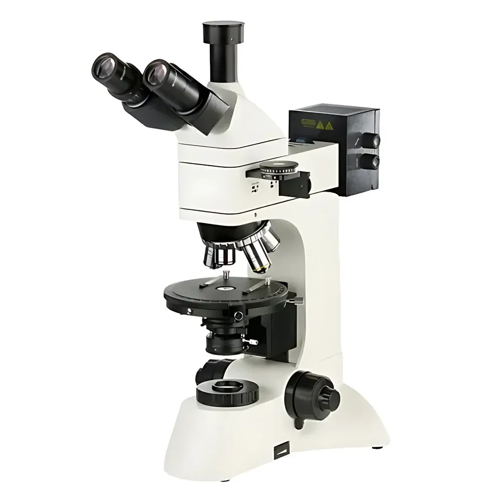
Betop Scientific XPL-3230 Polarizing Microscope
| Brand | Betop Scientific |
|---|---|
| Origin | Guangdong, China |
| Manufacturer Type | Direct Manufacturer |
| Model | XPL-3230 |
| Optical System | Infinity-Corrected |
| Objective Lenses | Strain-Free Plan Achromatic Polarizing Objectives (5X, 10X, 40X, 60X) |
| Eyepieces | Widefield WF10X (Φ22 mm), Divided Eyepiece 10X (0.1 mm/div) |
| Rotating Stage | 360° Graduated, Vernier Resolution 6′, Center-Adjustable with Lock |
| Focusing Mechanism | Coaxial Coarse/Fine Focus, Fine Adjustment Graduation 2 µm, Limit & Lock Function |
| Illumination | 6V/30W Halogen Lamp (Transmitted & Reflected), Brightness Adjustable |
| Polarizers | Rotatable Polarizer & Analyzer (0–360°), Graduated Scale with Vernier |
| Condenser | Abbe Condenser NA=1.25 with Iris Diaphragm |
| Compensation Plates | λ, λ/4, and Quartz Wedge |
| Trinocular Tube | 30° Inclined, 100% Light Path for Imaging |
| Power Supply | 85–265 V AC, 50/60 Hz |
Overview
The Betop Scientific XPL-3230 Polarizing Microscope is an advanced research-grade instrument engineered for quantitative birefringence analysis across materials science, geology, pharmaceutical development, and clinical pathology. It operates on the fundamental principle of polarized light interference—utilizing orthogonal linear polarization states (via a precisely aligned polarizer-analyzer pair) to reveal anisotropic optical properties in crystalline, fibrous, or stressed amorphous specimens. The microscope’s infinity-corrected optical architecture eliminates residual stress-induced artifacts in the imaging path, ensuring high-fidelity retardation measurements essential for identifying crystal symmetry, molecular orientation, and internal strain distribution. Designed for both transmitted-light (orthoscopic and conoscopic) and incident-light (reflected-polarized) observation modes, the XPL-3230 supports rigorous qualitative and semi-quantitative evaluation of birefringent features—including extinction angles, interference colors, and optic axis orientation—without compromising mechanical stability or ergonomic usability.
Key Features
- Infinity-corrected optical pathway with strain-free plan achromatic polarizing objectives (5×, 10×, 40×, 60×), each optimized for minimal intrinsic birefringence and long working distance (up to 26.1 mm at 5×).
- Widefield eyepieces (WF10×, Φ22 mm field number) and a calibrated divided eyepiece (0.1 mm per division) enable precise spatial referencing during crystallographic measurement.
- 30° inclined trinocular head with 100% light diversion capability—ensuring maximum photon throughput for low-light digital imaging applications including time-lapse birefringence monitoring.
- High-precision rotating mechanical stage (Φ150 mm) featuring 360° vernier scale (6 arcminute resolution), center-adjustable rotation axis, and dual-axis lock mechanism for repeatable angular positioning under load.
- Independent rotatable polarizer and analyzer, each equipped with engraved angular scales and fine vernier adjustment—critical for accurate extinction angle determination per ASTM E112 and ISO 9276-2 standards.
- Dual illumination system: 6 V/30 W halogen source for both Köhler-transmitted and episcopic reflected illumination, with continuous brightness control and integrated field/iris diaphragms for optimal contrast management.
- Standard compensation optics include full-wave (λ), quarter-wave (λ/4), and quartz wedge retardation plates—enabling determination of sign and magnitude of birefringence in unknown anisotropic samples per USP guidelines.
Sample Compatibility & Compliance
The XPL-3230 accommodates standard 26 mm × 76 mm glass slides and 12 mm diameter cover slips, supporting both thin-section petrographic mounts and thick-section metallurgical specimens up to 30 mm vertical clearance. Its modular design permits seamless integration of optional accessories—including five-position objective turrets, precision mechanical stages (30 mm × 25 mm travel), and C-mount CCD adapters compliant with IEEE 1394 and USB 2.0 protocols. The instrument meets electromagnetic compatibility requirements per IEC 61326-1 and electrical safety standards per IEC 61010-1. While not certified for GMP production environments, its stable optical calibration, traceable stage graduation, and reproducible polarizer alignment support GLP-compliant documentation workflows in academic, regulatory, and industrial QA/QC laboratories.
Software & Data Management
The XPL-3230 interfaces with third-party imaging platforms via standardized video (NTSC/PAL) and USB outputs. Compatible acquisition software—including open-source tools such as ImageJ/Fiji with polarized light plugins—supports annotation of extinction positions, interference color charts (Michel-Lévy), and birefringence quantification using calibrated retardation references. Optional DV-series digital cameras (DV-1 to DV-3) provide real-time analog output (380–520 TV lines) or progressive-scan digital capture (up to 10 MP resolution), all retaining timestamped metadata for audit trails. No proprietary firmware or vendor-locked drivers are required; raw image export formats (TIFF, BMP, AVI) ensure interoperability with LIMS and ELN systems used in pharmaceutical and geological repositories.
Applications
- Pharmaceutical solid-state characterization: polymorph identification, hydrate/anhydrate differentiation, and crystallinity assessment of active pharmaceutical ingredients (APIs) per ICH Q5A and USP .
- Geological thin-section analysis: optical mineralogy, twinning recognition, and optic sign determination in silicates, carbonates, and sulfides per ASTM D5776 and ISO 14688-1.
- Clinical crystallography: detection and morphological classification of monosodium urate (gout), calcium pyrophosphate (pseudogout), and oxalate crystals in synovial fluid per CLSI EP12-A2.
- Materials engineering: residual stress mapping in polymer films, fiber orientation analysis in composites, and phase segregation studies in liquid crystalline polymers.
- Cultural heritage science: pigment layer stratigraphy, binder identification in historical paintings, and degradation product analysis in mural plaster substrates.
FAQ
What types of samples can be observed in reflected-polarized mode?
The XPL-3230 supports opaque and semi-opaque specimens—including metallographic sections, sintered ceramics, and coated surfaces—using its dedicated episcopic illumination path and strain-free objectives optimized for high numerical aperture and minimal glare.
Is the microscope compatible with automated stage controllers or motorized focus units?
While the base configuration uses manual mechanical controls, the trinocular port and standardized mounting threads allow aftermarket integration with stepper-motorized XY stages and Z-axis focus drives meeting NEMA 17 specifications.
How is calibration verified for quantitative birefringence measurements?
Users perform periodic verification using NIST-traceable quartz reference slides (e.g., Berek or Soleil compensators) and certified interference color charts; angular scale accuracy is confirmed via autocollimation or laser interferometry against metrology-grade rotary encoders.
Can the XPL-3230 meet FDA 21 CFR Part 11 requirements for electronic records?
The microscope itself does not embed electronic record functionality; however, when paired with validated third-party imaging software featuring audit trail, electronic signature, and role-based access controls, it may be incorporated into Part 11–compliant workflows within broader laboratory quality systems.