Cairn Optosplit Dual-Channel Simultaneous Imaging Splitter
| Brand | Cairn |
|---|---|
| Origin | United Kingdom |
| Model | Optosplit |
| Component Type | Optical Beam-Splitting Assembly |
| Wavelength Separation | Dual-band, adjustable via rotating dichroic prism |
| Spatial Registration | Precise co-aligned imaging via fixed optical path difference |
| Field Selection | Optional variable-field iris for ROI definition and inter-channel crosstalk suppression |
| Compliance | Designed for integration into ISO 17025-compliant microscopy workflows |
| Interface | Standard C-mount and SM1-threaded optical mounts |
Overview
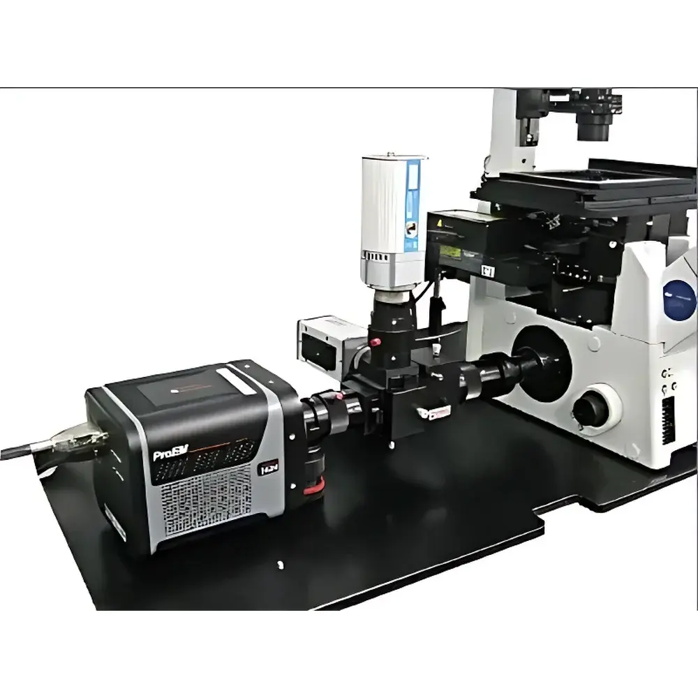
The Cairn Optosplit Dual-Channel Simultaneous Imaging Splitter is an optomechanical beam-splitting assembly engineered for high-fidelity, temporally synchronized acquisition of two spectrally distinct emission channels on a single scientific camera. Unlike sequential filter-wheel-based approaches—whose mechanical switching introduces temporal offsets exceeding tens to hundreds of milliseconds—the Optosplit employs a fixed-path, dichroic prism-based architecture that ensures true simultaneity (Δt < 1 µs) between the two output image planes. This eliminates motion-induced misregistration in live-cell or fast-dynamic experiments and preserves quantitative fidelity in ratiometric measurements. The device operates on the principle of spectral dispersion via a rotatable dichroic beamsplitter, enabling precise alignment of emission bands across a wide range of fluorophore pairs (e.g., CFP/YFP, GFP/mCherry, Alexa Fluor 488/Alexa Fluor 594). Its compact, all-metal housing provides thermal and mechanical stability essential for long-duration time-lapse acquisitions under vibration-sensitive conditions.
Key Features
- True simultaneous dual-channel imaging with sub-microsecond temporal synchronization
- Rotatable dichroic prism mount for precise, repeatable bandpass alignment without realignment of downstream optics
- Adjustable spatial separation between output image fields to accommodate pixel binning, sensor size, and microscope magnification
- Optional variable-field iris aperture for user-defined region-of-interest (ROI) selection and effective suppression of inter-channel optical crosstalk (typical isolation >99.3% at specified band edges)
- C-mount and SM1-threaded interfaces ensure compatibility with standard EMCCD, sCMOS, and scientific CMOS cameras and inverted/epifluorescence microscopes
- Optimized for minimal wavefront distortion: transmitted and reflected paths maintain <λ/8 RMS wavefront error across 400–750 nm
Sample Compatibility & Compliance
The Optosplit is routinely deployed in regulated and non-regulated life science laboratories performing quantitative fluorescence microscopy. It supports sample types ranging from fixed tissue sections and cultured adherent cells to suspended primary neurons and zebrafish embryos. Its optical design conforms to common requirements for GLP-compliant imaging workflows, including traceable alignment procedures and stable mechanical registration over temperature fluctuations (operating range: 15–30 °C). While the Optosplit itself is not a standalone measurement instrument, its integration into imaging systems intended for regulatory submissions (e.g., FDA 21 CFR Part 11–compliant platforms) is facilitated by its passive, calibration-free operation and absence of electronic control dependencies. It is compatible with ISO/IEC 17025-accredited calibration protocols when used with NIST-traceable reference standards for intensity linearity and spectral throughput verification.
Software & Data Management
The Optosplit requires no dedicated software or firmware—it functions as a hardware-level optical interface. Image data acquisition is fully managed by the host camera’s native SDK (e.g., Andor SDK, Hamamatsu DCAM, Photometrics PVCAM) or open-source frameworks such as Micro-Manager and PyMeasure. Post-acquisition, dual-channel frames are stored as side-by-side or top-bottom tiled TIFF stacks, preserving native bit depth and metadata. Common analysis pipelines—including ratiometric quantification (e.g., [Ca²⁺]i via Fura-2), FRET efficiency calculation using sensitized emission or acceptor photobleaching methods, and TIRF evanescent field depth profiling—leverage standard tools like Fiji/ImageJ, MATLAB, or Python-based libraries (e.g., scikit-image, pycro-manager). Audit trails for image acquisition parameters (exposure time, gain, ROI coordinates) are maintained within the camera’s metadata header, supporting traceability in GxP environments.
Applications
- Ion ratio imaging: Real-time quantification of intracellular ion concentrations (e.g., Ca²⁺, pH, Cl⁻) using dual-excitation or dual-emission ratiometric dyes
- Dual-label fluorescence imaging: Co-localization analysis of protein-protein interactions, organelle dynamics, and receptor trafficking without temporal interpolation artifacts
- Fluorescence resonance energy transfer (FRET): Accurate donor/acceptor intensity ratio acquisition for proximity-based molecular interaction studies
- Total internal reflection fluorescence (TIRF) microscopy: Simultaneous monitoring of membrane-proximal events and cytosolic references to normalize evanescent field variability
- Multi-spectral particle tracking: Concurrent trajectory mapping of spectrally encoded nanocarriers or quantum dots in complex media
FAQ
Does the Optosplit require external power or software drivers?
No. It is a purely passive optical component with no electronics, motors, or firmware.
Can it be used with confocal or light-sheet microscopes?
Yes—provided the system’s detection path delivers collimated or near-collimated beam geometry compatible with the Optosplit’s input NA specification (f/4 to f/8 recommended).
How is spectral crosstalk quantified and minimized?
Crosstalk is mitigated via high-performance dichroic coatings and the optional variable-field iris; typical values are validated using monochromatic light sources and spectroradiometric measurement per ISO 9288.
Is calibration documentation available for regulatory submissions?
Cairn provides optical performance test reports (including transmission curves, spatial separation tolerance, and wavefront error maps) upon request—suitable for inclusion in IQ/OQ documentation packages.
What camera sensor formats are supported?
All standard scientific sensors up to 22 mm diagonal (e.g., 2048 × 2048 sCMOS) are accommodated; custom spacers are available for larger formats.


