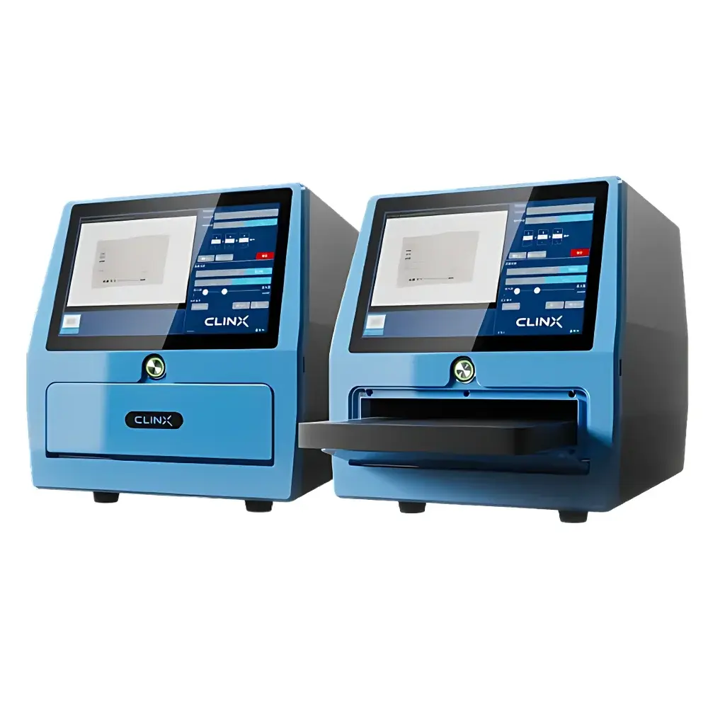
ChemiScope S7 Mini High-Sensitivity Chemiluminescence Imaging System
| Brand | Clinx |
|---|---|
| Origin | Shanghai, China |
| Model | ChemiScope S7 |
| Instrument Type | Chemiluminescence Gel Imaging System |
| CCD Resolution | 6.0 MP |
| Bit Depth | 16-bit |
| Dynamic Range | 4.8 OD |
| Effective Sensor Resolution | 1440 × 1004 pixels |
| Pixel Size | 96 µm × 96 µm |
| Max Imaging Area | 145 mm × 110 mm |
| Exposure Modes | Auto, Manual, Sequential |
| Exposure Time Range | 0.1 s – 200 s |
Overview
The ChemiScope S7 Mini High-Sensitivity Chemiluminescence Imaging System is a compact, integrated benchtop imager engineered for quantitative detection of chemiluminescent signals in nucleic acid and protein electrophoresis applications. It operates on the principle of photon-counting imaging using a cooled scientific-grade CCD sensor optimized for low-light signal acquisition. Unlike conventional gel documentation systems relying on visible-light transillumination or UV excitation, the ChemiScope S7 captures emitted photons directly from enzymatic (e.g., HRP- or AP-mediated) or non-enzymatic chemiluminescent reactions—enabling detection at sub-femtogram levels of target analytes. Its optical architecture incorporates a high-transmission lens system, thermoelectrically stabilized sensor cooling (−25 °C below ambient), and precision light-tight enclosure design to minimize dark current and stray light interference. This configuration ensures reproducible quantification across repeated exposures and supports compliance-driven workflows in academic research, biopharmaceutical QC, and molecular diagnostics laboratories.
Key Features
- Photon-lossless imaging architecture with ultra-low read noise (65% at 520 nm), enabling detection of weak chemiluminescent signals without signal amplification artifacts.
- True-color molecular weight marker overlay functionality: simultaneous acquisition and registration of colorimetric reference lanes alongside monochrome chemiluminescent signals, preserving spatial fidelity for accurate band alignment and annotation.
- Intelligent exposure control algorithm that dynamically adjusts integration time and gain based on real-time signal saturation analysis—eliminating iterative trial-and-error exposure optimization.
- Integrated embedded computing platform: self-contained Windows-based imaging station with onboard SSD storage, eliminating dependency on external PCs and reducing electromagnetic interference risks in shared lab environments.
- Compact footprint (W × D × H: 380 × 360 × 420 mm) designed for space-constrained biosafety cabinets or laminar flow hoods without compromising optical path length or field uniformity.
Sample Compatibility & Compliance
The ChemiScope S7 accommodates standard vertical and horizontal electrophoresis gels (up to 145 mm × 110 mm), blots (PVDF, nitrocellulose, nylon), autoradiography films, and microplate-based chemiluminescent assays. It supports common detection chemistries including Luminol/Peroxide (HRP), CSPD/CDP-Star (AP), and acridan-based substrates. All image metadata—including exposure parameters, timestamp, user ID, instrument serial number, and calibration status—is embedded into TIFF and PNG exports in EXIF-compliant format. The system’s software architecture adheres to ALCOA+ principles (Attributable, Legible, Contemporaneous, Original, Accurate, Complete, Consistent, Enduring, Available) and supports audit trail generation required under GLP and GMP frameworks. While not FDA 21 CFR Part 11–certified out-of-the-box, the system can be validated per internal SOPs for regulated environments when deployed with controlled access protocols and electronic signature modules.
Software & Data Management
The ChemiScope S7 ships with two complementary software suites: ChemiCapture (onboard acquisition) and ChemiAnalyze (desktop analysis). ChemiCapture provides intuitive workflow management for single-shot, time-series, and multi-channel acquisitions—with automatic alignment of chemiluminescent and color reference images. All raw frames are saved in lossless 16-bit TIFF format with full metadata retention. ChemiAnalyze delivers robust post-acquisition processing, including background subtraction (rolling ball algorithm), lane/strip auto-detection with manual override capability, band intensity quantification normalized to internal controls or total lane density, and statistical reporting (CV%, fold-change, p-value estimation via t-test). Export options include CSV, Excel (.xlsx), PDF reports with embedded graphs, and publication-ready PNG/TIFF with DPI scaling up to 600 dpi. Data integrity is enforced through write-protected archive folders and optional network drive synchronization.
Applications
- Western blot quantification of low-abundance phosphoproteins, cytokines, and transcription factors.
- High-sensitivity Northern/Southern blot detection of rare mRNA isoforms or methylated DNA fragments.
- Validation of CRISPR-Cas9 editing efficiency via chemiluminescent probe hybridization.
- Quality control of recombinant antibody batches using HRP-conjugated secondary detection.
- Time-resolved kinetic assays monitoring enzyme activity or ligand-receptor binding via luminogenic substrates.
- Teaching laboratory implementation where reproducibility, ease of use, and minimal infrastructure requirements are critical success factors.
FAQ
What is the effective pixel resolution used for quantitative analysis?
The active imaging sensor delivers 1440 × 1004 pixels at 16-bit depth; binning modes are not applied during default acquisition to preserve spatial resolution for precise band localization.
Does the system support multiplex chemiluminescent detection?
Yes—sequential imaging with spectral filtering (optional add-on filter wheel) enables dual-probe detection using spectrally distinct substrates (e.g., near-infrared vs. blue-emitting luminophores), provided emission peaks are resolvable by the native lens transmission profile.
Can exposure settings be scripted for unattended overnight runs?
Absolutely—the ChemiCapture software supports batch protocol scheduling with conditional triggers (e.g., “acquire if signal > threshold X”), automatic file naming conventions, and error logging for long-duration kinetics experiments.
Is service calibration traceable to NIST standards?
Yes—factory calibration includes uniformity correction maps and sensitivity reference checks against certified luminance standards (NIST SRM 2242), with recalibration intervals recommended annually or after 5000 operational hours.
How is data security handled in shared-lab deployments?
User accounts are managed via Windows Active Directory integration; role-based permissions restrict access to acquisition modules, analysis tools, and export functions—ensuring separation of duties between operators, analysts, and administrators.
Related Products