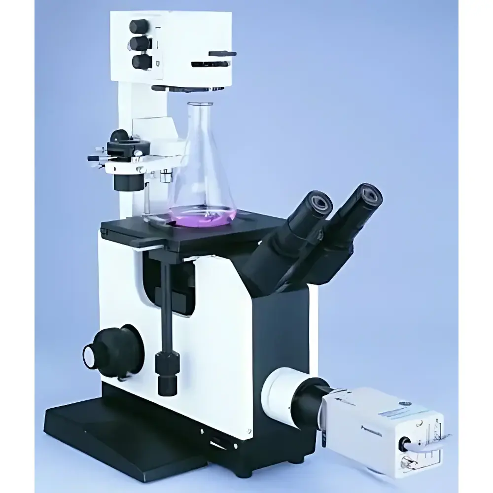
Chongqing Optics XDS-1B Inverted Biological Microscope
| Brand | Chongqing Optics |
|---|---|
| Origin | Chongqing, China |
| Model | XDS-1B |
| Total Magnification | 100×–640× |
| Illumination | Built-in 6 V / 20 W halogen lamp with adjustable brightness and filter wheel |
| Focus Mechanism | Coaxial coarse/fine focus with 20 mm travel, fine focus graduation 0.02 mm, upper-limit stop & locking mechanism |
| Eyepiece Tube | Dual-port 45° inclined binocular tube, interpupillary adjustment 55–75 mm, diopter correction |
| Objective Turret | 4-position, centering precision turret with ball-bearing rotation and anti-fungal coating |
| Stage | Rectangular mechanical stage (155 × 180 mm), X–Y travel 50 × 75 mm |
| Condenser | Long-working-distance condenser, W.D. = 55.2 mm, NA = 0.3 |
| Objectives | LPL Plan Achromatic Long Working Distance Objectives — LPL10×/0.25 (W.D. 7.9 mm), LPL25×/0.40 (W.D. 5 mm), LPL40×/0.65 (W.D. 3 mm) |
| Phase Contrast | Optional Ph25× objective (W.D. 5 mm) with 25× phase ring slider |
| Eyepieces | Wide-field WF10×/20, WF16×/14, WF10× reticle/18, 11× centering eyepiece |
| Compliance | ISO 9001:2015, ISO 14001:2015, CE (Low Voltage Directive 2014/35/EU), Medical Device Registration No.: Yu Food & Drug Admin Med Cert (Approval) No. 2007-2220053 |
Overview
The Chongqing Optics XDS-1B Inverted Biological Microscope is engineered for routine and advanced live-cell observation in academic laboratories, pharmaceutical QC environments, and biotechnology R&D facilities. Its inverted optical architecture places the objective lenses beneath the specimen stage—enabling direct visualization of adherent or suspension cells cultured in standard Petri dishes, multi-well plates, and flasks without specimen inversion or mounting. The system employs Köhler illumination with a built-in 6 V / 20 W halogen source, optimized for stable thermal output and consistent color temperature across intensity settings. Optical design follows classical finite-conjugate principles with plan achromatic long-working-distance (LWD) objectives, minimizing spherical and chromatic aberrations while maximizing flatness of field and edge-to-edge resolution. This configuration supports non-invasive, label-free monitoring of cellular morphology, motility, confluence, and mitotic progression—particularly valuable in time-lapse imaging workflows where minimal phototoxicity and mechanical stability are critical.
Key Features
- Long-working-distance plan achromatic objectives (10×, 25×, 40×) with working distances up to 7.9 mm—accommodating thick culture vessels and accessories such as incubation chambers or micromanipulators.
- Coaxial coarse/fine focusing mechanism with 20 mm vertical travel, calibrated fine-focus scale (0.02 mm per division), mechanical upper-limit stop, and focus-locking capability for repeatable Z-positioning during multi-site acquisition.
- Rectangular mechanical stage (155 × 180 mm) with 50 × 75 mm travel range and low-profile controls—designed for ergonomic access during extended observation sessions.
- 45° inclined dual-port binocular head with interpupillary adjustment (55–75 mm), individual diopter compensation, and optional photo-port for simultaneous visual inspection and image capture.
- Phase contrast compatibility via dedicated LPL Ph25× and optional LPL Ph40× objectives, paired with interchangeable annular diaphragms—enabling high-contrast visualization of unstained, transparent specimens including primary neurons, fibroblasts, and embryoid bodies.
- Integrated filter wheel accommodating neutral density, green interference, and blue excitation filters—facilitating basic fluorescence screening when paired with external LED or mercury lamp sources (not included).
Sample Compatibility & Compliance
The XDS-1B accommodates standard cell culture formats—including 35 mm, 60 mm, and 100 mm Petri dishes; T-25, T-75, and T-175 flasks; and 6-, 12-, 24-, 48-, and 96-well plates—without requiring sample transfer or coverslip mounting. Its 55.2 mm working distance condenser (NA 0.3) ensures uniform illumination across vessel bases, even under ambient lab lighting conditions. Regulatory compliance includes ISO 9001:2015 (Quality Management Systems), ISO 14001:2015 (Environmental Management), and CE marking under the EU Low Voltage Directive (2014/35/EU). As a Class IIa medical device registered with the Chongqing Municipal Medical Products Administration (Registration No. Yu Food & Drug Admin Med Cert [Approval] No. 2007-2220053), it meets essential requirements for safety, electromagnetic compatibility, and biocompatibility of user-facing components.
Software & Data Management
While the XDS-1B operates as an analog optical platform, its dual-port design enables seamless integration with third-party digital imaging systems. Compatible accessories include C-mount adapters for DSLR/mirrorless cameras, USB 3.0 CMOS sensors (e.g., 5 MP–12 MP monochrome/color modules), and analog video outputs for legacy CCTV or frame-grabber acquisition. When paired with validated software packages—such as ImageJ/Fiji (open-source), NIS-Elements (Nikon), or CellSens (Olympus)—the system supports time-series capture, Z-stack reconstruction, region-of-interest (ROI) intensity profiling, and basic morphometric analysis. Audit trail functionality, user access control, and electronic signature support require integration with compliant laboratory information management systems (LIMS) or ELN platforms meeting FDA 21 CFR Part 11 criteria.
Applications
- Monitoring proliferation and confluence of mammalian cell lines (e.g., HEK293, HeLa, CHO-K1) during bioprocess development.
- Assessing viability and morphology of primary isolates (e.g., hepatocytes, cardiomyocytes, neural stem cells) under controlled environmental conditions.
- Performing manual cell counting and colony formation assays using reticle eyepieces and calibrated stage micrometers.
- Supporting microinjection, patch-clamp setup alignment, and micromanipulator-guided subcellular targeting.
- Validating sterilization efficacy and contamination presence in media or reagent batches via direct visual inspection of microbial growth.
- Teaching core microscopy principles—including Köhler illumination, phase contrast theory, depth of field estimation, and resolution limits—in undergraduate life science curricula.
FAQ
Is the XDS-1B suitable for fluorescence imaging?
The base model does not include epi-illumination or filter cubes. However, fluorescence observation is feasible when coupled with an external LED or mercury lamp source and appropriate excitation/emission filter sets mounted at the photo-port or condenser aperture.
Can I upgrade to motorized focus or automated stage control?
The XDS-1B features a manually operated mechanical stage and focus system. Motorized upgrades are not factory-supported; integration requires third-party actuators compatible with standard M42 or C-mount interfaces and external controller hardware.
What is the maximum thickness of culture dish supported?
With the 55.2 mm working distance condenser and LPL10× objective (7.9 mm W.D.), the system accommodates dishes up to ~12 mm total height—including lid clearance—provided the bottom surface remains optically flat and free of scratches or debris.
Does the microscope meet GLP or GMP documentation requirements?
As supplied, the XDS-1B includes calibration certificates for optical alignment and illumination uniformity. Full GLP/GMP compliance requires additional validation protocols (IQ/OQ/PQ), maintenance logs, and traceable calibration against NIST-traceable standards—typically implemented through institutional QA departments.
Are replacement parts and service available outside China?
Chongqing Optics maintains an international distributor network; technical documentation, spare objectives, eyepieces, bulbs, and condenser components are available through authorized partners in North America, EMEA, and APAC regions—with lead times varying by component availability and shipping logistics.


