Countstar Spica Real-Time Intelligent Live-Cell Imaging and Analysis System
| Brand | Countstar |
|---|---|
| Origin | Shanghai, China |
| Model | Countstar Spica |
| Type | Label-Free Live-Cell Imaging System |
| Optical Configurations | Spica M1 (7.5×/10×/15× |
| 3-channel fluorescence | DAPI/GFP/RFP |
| 4-channel fluorescence | DAPI/GFP/RFP/APC |
| Environmental Integration | Incubator-compatible (37 °C, 5% CO₂, humidity control) |
| Software | AI-powered analysis platform with ≥12 pre-validated application modules, FDA 21 CFR Part 11–compliant audit trail (optional), GLP/GMP-ready data export (CSV, TIFF, JSON, PDF) |
Overview
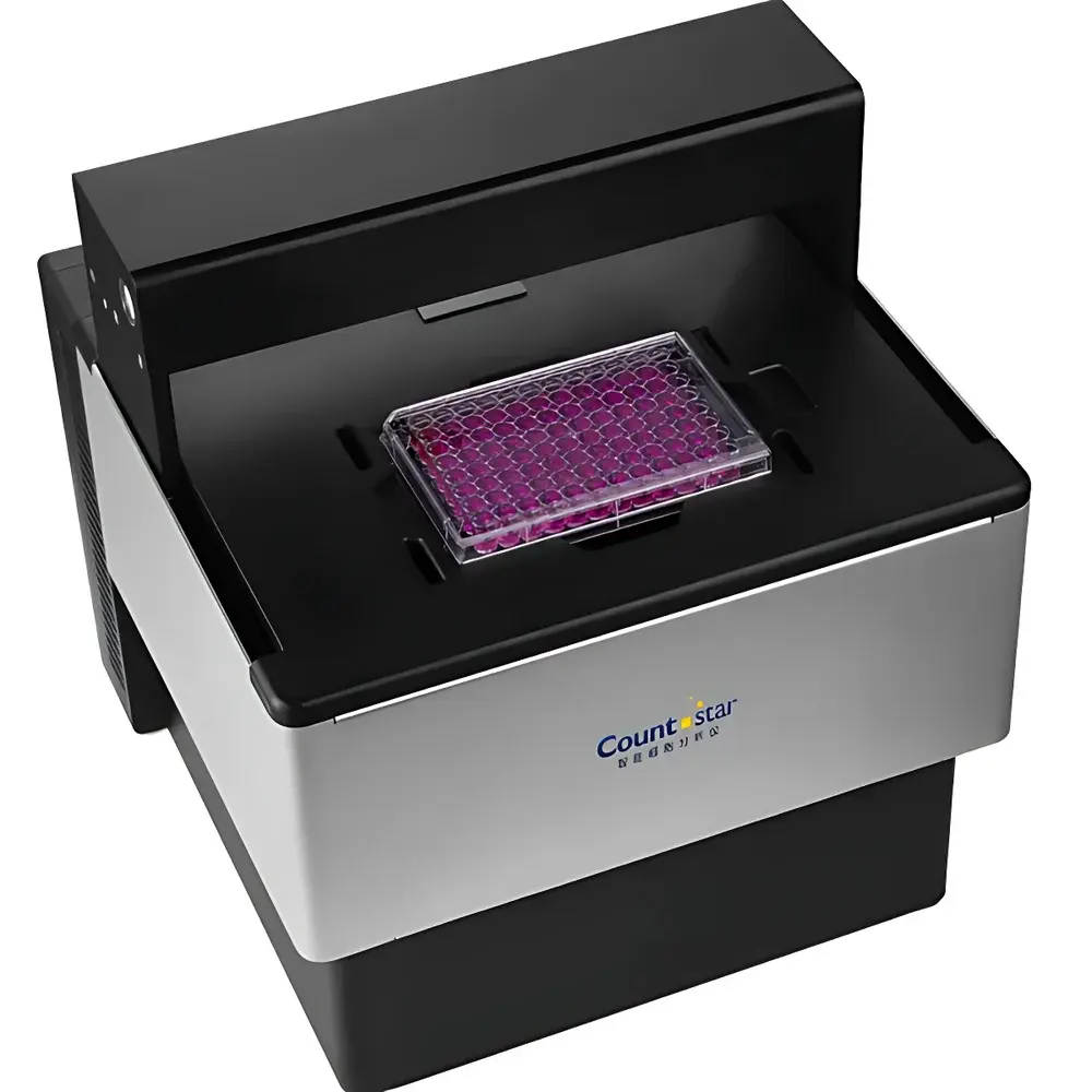
The Countstar Spica Real-Time Intelligent Live-Cell Imaging and Analysis System is an incubator-integrated, label-free quantitative imaging platform engineered for longitudinal, non-invasive monitoring of living cells under physiologically relevant conditions. Leveraging high-resolution wide-field microscopy combined with AI-driven image segmentation and temporal tracking algorithms, the Spica system captures dynamic cellular behaviors—including proliferation, migration, morphology changes, spheroid growth, and organoid development—without requiring exogenous dyes or genetic modification. Its core architecture supports both endpoint and time-lapse acquisition across standard multiwell plates (6–384-well), microfluidic chips, and custom substrates. Designed for seamless integration into regulated laboratory environments, the system operates continuously inside standard CO₂ incubators, maintaining precise thermal (±0.3 °C), gaseous (5% CO₂ ±0.2%), and humidity control throughout extended experiments lasting days to weeks.
Key Features
- Incubator-compatible hardware design with sealed optical path and vibration-dampened stage, enabling stable long-term imaging without compromising sterility or environmental fidelity
- Dual-platform configuration: Spica M1 optimized for high-content single-plate studies with micrometer-precision XYZ motorized stage and laser-assisted auto-focus; Spica M6 engineered for parallel multi-plate throughput with six independent plate positions and synchronized multi-channel acquisition
- Multi-magnification optics: M1 offers 7.5×/10×/15× objectives with integrated brightfield and three-fluorescence channels (DAPI/GFP/RFP); M6 provides 4×/10×/20× objectives with four-fluorescence capability (DAPI/GFP/RFP/APC) and ultra-low-noise EMCCD/sCMOS sensor support
- Z-stack acquisition engine with adaptive focus mapping and rapid layer scanning—capable of acquiring 20 optical sections per well in ≤8 minutes across a full 96-well plate
- Real-time AI inference pipeline trained on >50,000 annotated cell images, delivering sub-pixel segmentation accuracy and frame-to-frame object association with minimal user calibration
- FDA 21 CFR Part 11–ready software architecture featuring electronic signatures, immutable audit trails, role-based access control, and automated backup protocols compliant with ISO/IEC 17025 and GLP documentation standards
Sample Compatibility & Compliance
The Spica system accommodates standard tissue-culture-treated plates (including black-wall, clear-bottom, and glass-bottom variants), chamber slides, Lab-Tek II chambers, and hydrogel-embedded 3D cultures. It supports label-free phase contrast, differential interference contrast (DIC), and fluorescence modalities without phototoxicity escalation—validated per ISO 10993-5 for cytocompatibility during continuous illumination. All optical components meet RoHS and CE directives. System validation documentation—including IQ/OQ/PQ templates, traceable calibration certificates for stage positioning (±0.5 µm repeatability), and fluorescence intensity linearity reports (R² > 0.999 over 4-log dynamic range)—is provided for GxP-regulated workflows.
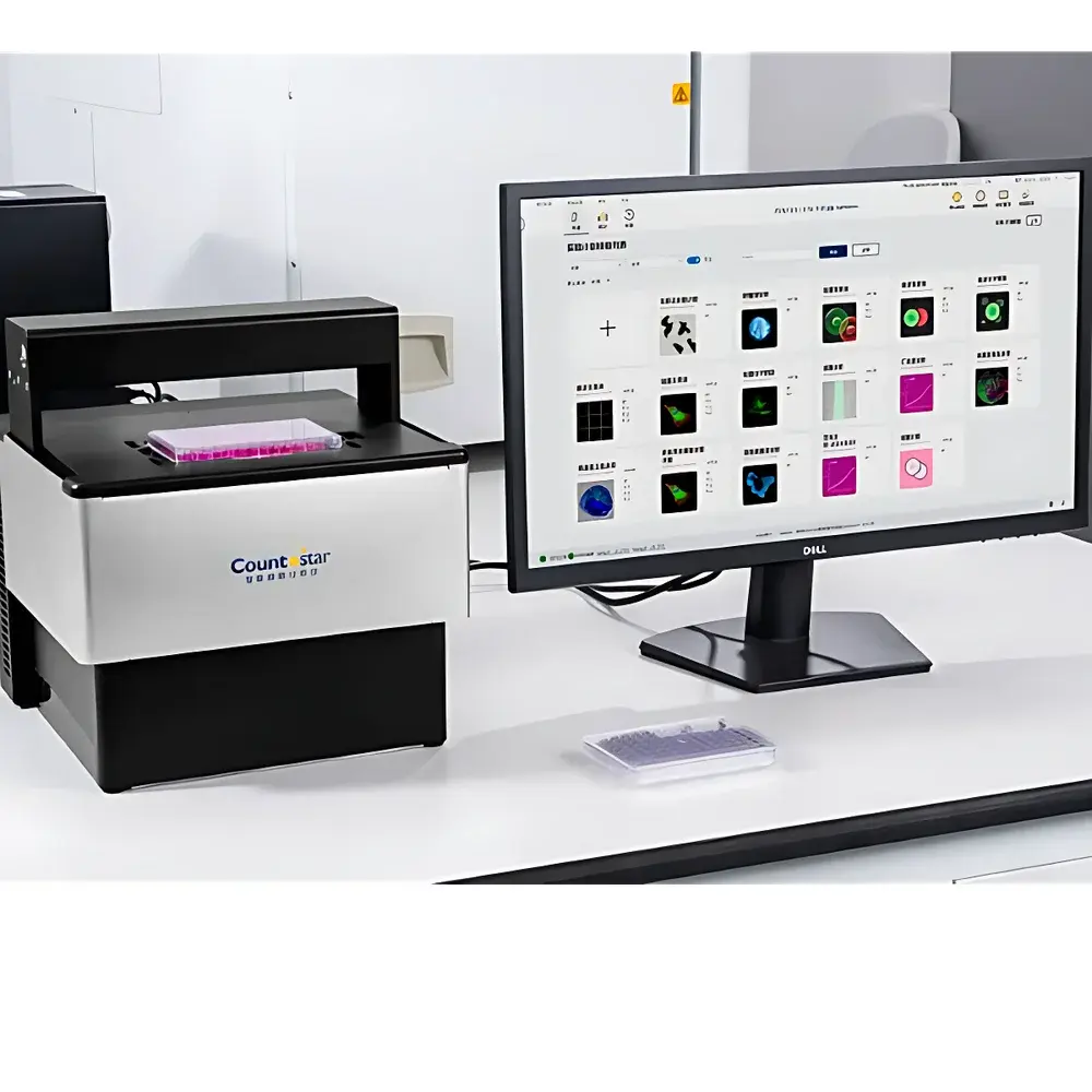
Software & Data Management
The Countstar Spica Analysis Suite v4.x is a modular, open-API platform supporting local deployment, networked lab server integration, and secure cloud synchronization (AES-256 encrypted). Core capabilities include: twelve pre-validated AI analysis templates (confluence, wound healing, spheroid area/volume, organoid branching, apoptosis kinetics, cytotoxicity dose-response, phagocytosis index, single-cell trajectory mapping, etc.); customizable BioApps framework allowing Python-based algorithm injection and parameter optimization; automated curve generation (e.g., growth kinetics, IC₅₀/EC₅₀ fitting using four-parameter logistic regression); and FCS (Fluorescence Correlation Spectroscopy) module for diffusion coefficient and molecular brightness quantification. Raw data (16-bit TIFF stacks), processed metadata (JSON), and publication-ready figures (SVG/PDF) are exportable with embedded MIAME-compliant experimental annotations.
Applications
The Spica platform serves as a primary tool in academic, biopharmaceutical, and contract research laboratories for applications including—but not limited to—real-time assessment of compound-induced cytostatic/cytotoxic effects (per ASTM E2903-21), 3D tumor spheroid drug penetration modeling, iPSC-derived organoid maturation profiling, CRISPR-edited lineage tracing, immune cell–target cell interaction kinetics, and high-content screening of autophagy or ferroptosis modulators. Its label-free capability enables longitudinal studies where fluorescent labeling would otherwise perturb signaling pathways or introduce batch variability—particularly critical in stem cell differentiation assays and primary neuron culture monitoring.
FAQ
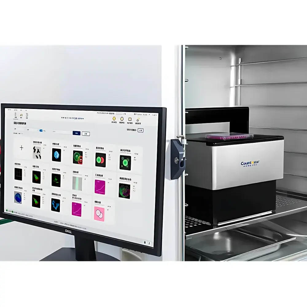
Can the Spica system operate inside a standard humidified CO₂ incubator?
Yes. The Spica M1 and M6 models are fully rated for continuous operation at 37 °C, 5% CO₂, and ≥95% relative humidity. All electronics are conformally coated, and optical paths are hermetically sealed against condensation.
Is the AI analysis module validated for regulatory submissions?
The core AI segmentation engine has undergone analytical verification per ICH Q5E and USP , with documented specificity (>98.2%), sensitivity (≥96.7%), and inter-operator reproducibility (CV < 3.1% across n=12 operators). Full validation packages are available upon request.
Does the system support third-party microscope objectives or custom filters?
Yes. The Spica optical interface complies with RMS (Royal Microscopical Society) thread standards and accepts C-mount filter cubes and infinity-corrected objectives from major OEMs (Olympus, Nikon, Zeiss, Leica) via optional adapter kits.
How is data integrity ensured during extended time-lapse experiments?
Each acquired frame is timestamped with NTP-synchronized UTC, checksum-verified upon write, and redundantly stored across dual SSD arrays. Power-fail recovery ensures no frame loss—even during unexpected outages.
Can analysis parameters be re-applied to raw data after experiment completion?
Absolutely. All raw image stacks are preserved in native format. Users may re-run any AI template—modified or unmodified—with full version history logging, eliminating the need for repeat acquisitions.
Related Products


