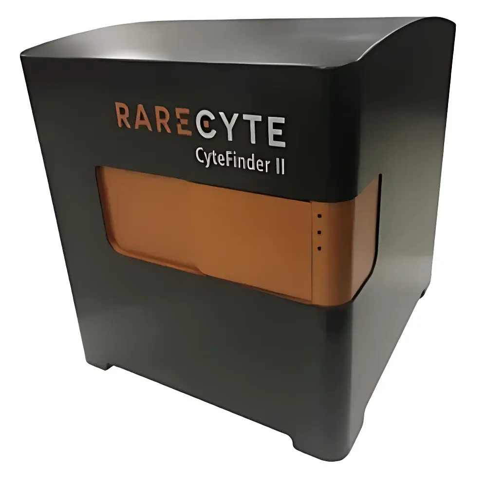
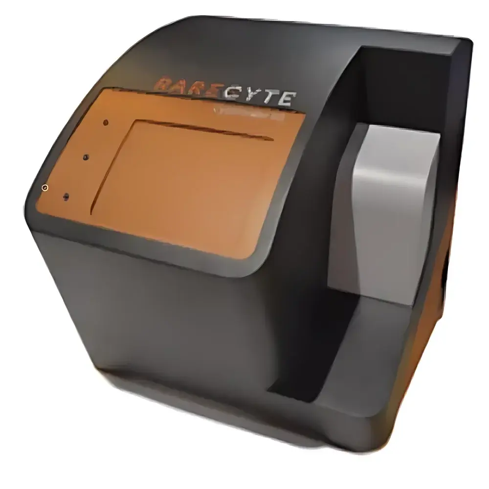
CyteFinder II Single-Cell Analysis Platform for CAR-T & Stem Cell Therapeutics
| Origin | USA |
|---|---|
| Manufacturer Type | Authorized Distributor |
| Origin Category | Imported |
| Model | CyteFinder II |
| Pricing | Available Upon Request |
Overview
The CyteFinder II is a high-throughput, whole-slide imaging and single-cell analysis platform engineered for translational research and clinical development in advanced cell therapies—including chimeric antigen receptor T-cell (CAR-T) manufacturing, stem cell characterization, and liquid biopsy–based diagnostics. Built upon a robust optical architecture combining wide-field fluorescence microscopy with precision motorized stage control, the system enables automated, quantitative detection and spatial profiling of ultra-rare cells at subcellular resolution. Its core measurement principle relies on multi-channel spectral separation (7-color fluorescence), AI-augmented image segmentation, and deterministic physical microdissection—ensuring minimal sample perturbation while preserving nucleic acid integrity for downstream NGS, qPCR, or functional assays. Designed for laboratories operating under GLP-compliant workflows and preparing for regulatory submissions (e.g., FDA IND/BLA dossiers), the platform supports traceable, audit-ready data acquisition aligned with ISO 13485–informed quality management systems.
Key Features
- Seven-channel simultaneous fluorescence imaging with high quantum efficiency sCMOS detection and customizable spectral filter sets (360–750 nm range)
- AI-powered rare-cell detection engine trained on >10,000 annotated CTC and immune cell morphologies; achieves ≥92% sensitivity and ≥88% specificity in spiked blood matrix validation studies
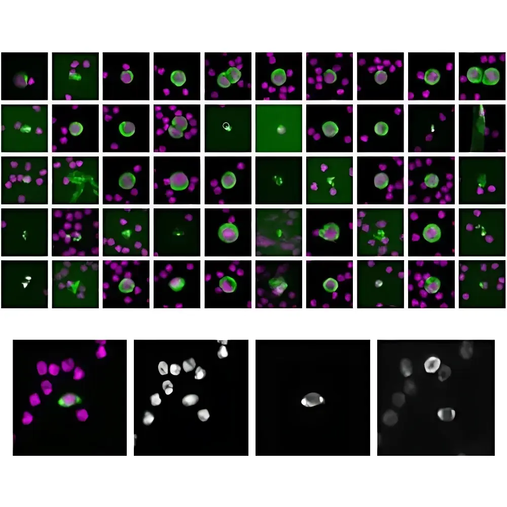
- Integrated CytePicker® module for contact-free, pressure-controlled microaspiration of individual live or fixed cells—enabling retrieval of intact, viable cells for clonal expansion, scRNA-seq, or functional phenotyping
- Whole-tissue section scanning capability up to 150 mm × 150 mm slides with <1 µm/pixel resolution in both brightfield and multiplexed fluorescence modes
- Automated tissue microregion isolation using laser-assisted microdissection (LMD) with real-time feedback and coordinate-mapped ROI export for spatial transcriptomics integration
- Modular hardware architecture supporting optional upgrades: environmental chamber (37°C, 5% CO₂), time-lapse imaging, and high-speed Z-stack acquisition (up to 50 µm/sec)
Sample Compatibility & Compliance
The CyteFinder II accepts standard glass slides (1 x 3 inches), cytospin preparations, membrane filters (e.g., ISET®, ScreenCell®), and FFPE/frozen tissue sections (4–50 µm thickness). It accommodates whole blood, PBMC isolates, bone marrow aspirates, CSF, pleural effusions, and dissociated organoids. All image acquisition and analysis modules comply with 21 CFR Part 11 requirements when deployed with validated software configuration (v4.2+), including electronic signatures, role-based access control, and immutable audit trails. The system meets ISO/IEC 17025 criteria for analytical method validation in clinical laboratory settings and supports documentation packages for CAP/CLIA accreditation readiness.
Software & Data Management
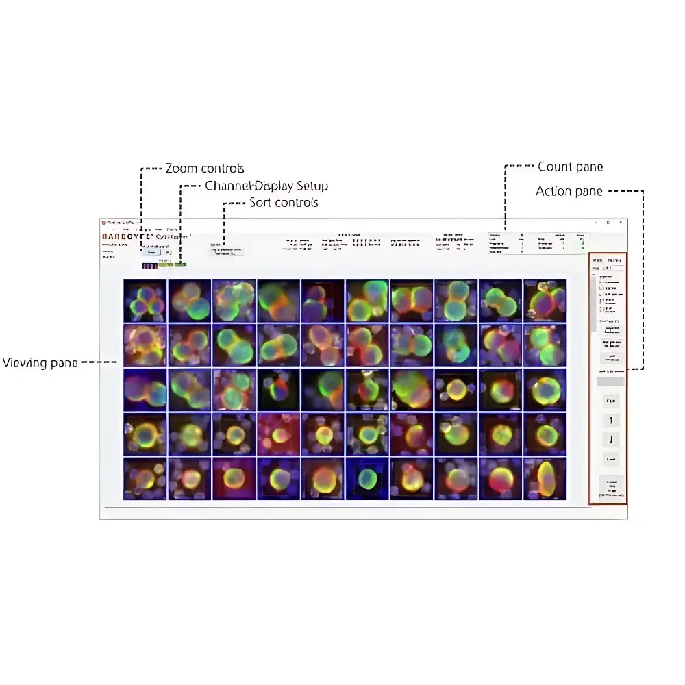
CyteFinder Studio v5.1 provides an integrated environment for acquisition, annotation, quantification, and metadata-driven reporting. The software features DICOM-SR compliant export, HL7/FHIR interface support for LIS/PACS integration, and native compatibility with common bioinformatics pipelines (e.g., Seurat, CellxGene, Loupe Browser). Image datasets are stored in vendor-agnostic OME-TIFF format with embedded OMERO metadata standards. Batch processing workflows—including cell classification, marker colocalization scoring, and spatial neighborhood analysis—are scriptable via Python API. All user actions, parameter changes, and result exports are logged with timestamped, user-identifiable entries meeting ALCOA+ principles.
Applications
- Liquid biopsy development: enumeration and molecular profiling of circulating tumor cells (CTCs), fetal nucleated red blood cells (fnRBCs), and circulating fetal cells (CFCs) from maternal peripheral blood
- CAR-T and TCR-T therapy QC: viability assessment, transduction efficiency verification, and phenotypic stability monitoring across manufacturing batches
- Stem cell potency assays: identification and isolation of pluripotent or lineage-committed progenitors from heterogeneous differentiation cultures
- Tumor immunology: spatial mapping of T-cell infiltration density, PD-1/PD-L1 co-expression patterns, and tertiary lymphoid structure (TLS) quantification in solid tumor microenvironments
- Immunophenotyping of rare PBMC subsets: detection of exhausted CD8⁺ T cells, regulatory T cells (Tregs), dendritic cell subsets, and antigen-presenting B cells
- Non-invasive prenatal testing (NIPT): isolation and genomic analysis of intact fetal cells for aneuploidy and monogenic disorder screening
- Single-cell multi-omics: coupling retrieved cells with targeted DNA methylation, full-length cDNA amplification, or ATAC-seq library preparation
FAQ
Does the CyteFinder II support live-cell imaging and recovery for functional assays?
Yes—the system includes optional environmental control modules and low-phototoxicity illumination protocols enabling long-term time-lapse imaging and subsequent retrieval of metabolically active cells for culture or electrophysiology.
Can the platform integrate with existing LIMS or hospital information systems?
Yes—via configurable HL7 v2.x and FHIR R4 interfaces, supporting bidirectional data exchange for specimen tracking, result reporting, and audit log synchronization.
Is the AI model retrainable with user-specific cell morphology data?
Yes—CyteFinder Studio includes a supervised training mode compatible with user-provided ground-truth annotations, allowing customization for novel biomarkers or species-specific cell types.
What regulatory documentation is provided for GMP-aligned operations?
A complete validation package is available, including IQ/OQ/PQ protocols, software design specifications, cybersecurity risk assessment (per IEC 62304), and 21 CFR Part 11 implementation guide.
How does the system ensure reproducibility across operators and sites?
Through standardized acquisition templates, instrument calibration certificates traceable to NIST standards, and inter-site concordance studies conducted per CLSI EP09-A3 guidelines.