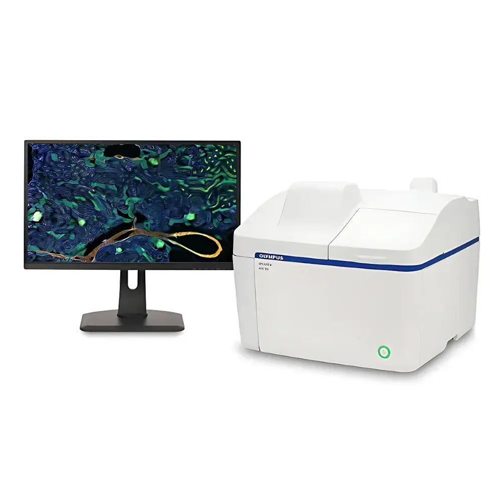
Evident APX100 Desktop Fluorescence Microscope
| Brand | Evident (formerly Olympus) |
|---|---|
| Origin | Japan |
| Manufacturer Type | Original Equipment Manufacturer (OEM) |
| Product Category | Imported Instrument |
| Model | APX100 |
| Configuration | Upright and Inverted Integrated Platform |
| Medical Device Classification | Non-Medical Device |
| Instrument Class | Research-Grade Fluorescence Microscope |
| Imaging Mode | Widefield Fluorescence + Gradient Contrast (GC) Transmitted Light |
| Software Interface | Intuitive, Workflow-Centric GUI with Automated Folder Structuring |
| Navigation System | Smart Sample Navigation with 0.07× Macro Overview Lens |
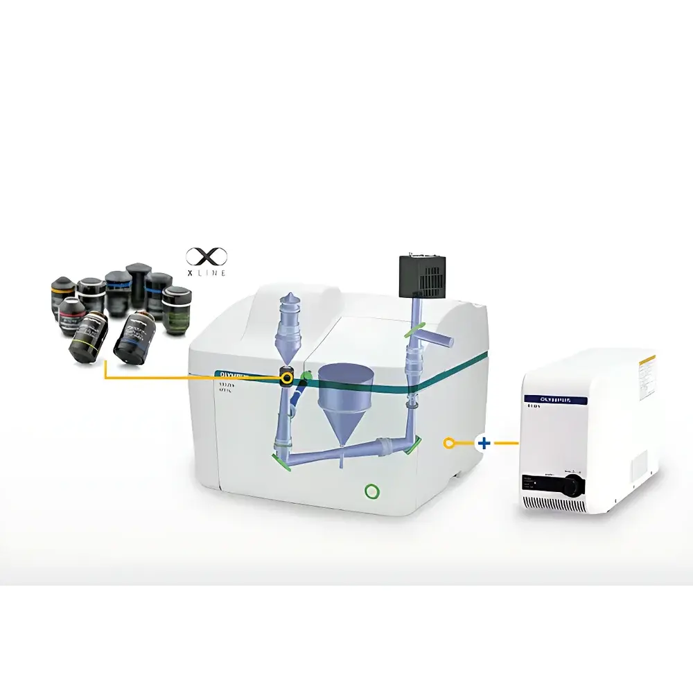
| Optical Path | Dual-Path (Fluorescence & GC) |
| Spectral Range | DAPI to Cy7 (350–800 nm) |
| Objective Compatibility | UPLXAPO Series (e.g., 4×, 20× APO), Standard Coverslip-Corrected Objectives |
Overview
The Evident APX100 Desktop Fluorescence Microscope is a compact, integrated upright/inverted research-grade imaging platform engineered for high-fidelity widefield fluorescence and label-free transmitted-light microscopy in life science laboratories. Designed around a rigid, vibration-damped enclosure, the APX100 eliminates environmental dependency—operating reliably under ambient lighting without requiring darkroom conditions. Its core optical architecture leverages Evident’s legacy in precision optics, incorporating proprietary Gradient Contrast (GC) technology—a non-invasive, filter-based transmitted-light method that generates pseudo-3D contrast in unstained live cells and tissue sections without phase rings, special slides, or specialized objectives. Combined with high-numerical-aperture apochromatic objectives and uniform LED illumination across excitation bands (350–800 nm), the system delivers quantitative fluorescence intensity linearity, accurate color fidelity in brightfield, and subcellular resolution in both fixed and live specimens.
Key Features
- Integrated upright/inverted configuration enables seamless transition between adherent cell cultures (inverted mode) and tissue sections or multiwell plates (upright mode) without repositioning samples.
- Smart Sample Navigation system employs a dedicated 0.07× macro lens to capture full-sample overviews in <1 second; users click any region of interest to auto-center and refocus at target magnification within 10 seconds.
- Automated data structuring: software generates time-stamped, experiment-specific subfolders upon acquisition—grouping raw images, metadata (objective, exposure, filter set), and processed outputs for audit-ready traceability.
- Gradient Contrast (GC) optical module provides high-contrast, depth-perceptive brightfield imaging of transparent, unstained biological specimens—compatible with standard glass-bottom dishes and plasticware.
- Multi-spectral fluorescence support from DAPI through Cy7 ensures compatibility with common nuclear, cytoskeletal, membrane, and synaptic markers—including Hoechst, GFAP, MAP2, Calbindin, MBP, DiO, DiI, and DiD.
- LED-based illumination with stable intensity control (<0.5% drift/hour) and programmable exposure sequencing enables reproducible time-lapse experiments up to 18+ hours with minimal phototoxicity.
Sample Compatibility & Compliance
The APX100 accommodates standard life science sample formats: 35 mm–100 mm Petri dishes, 6–96-well plates, glass slides, and chambered coverslips. Its mechanical stage supports XYZ motorization (optional) and precise Z-stepping for z-stack acquisition. The system complies with ISO 9001 manufacturing standards and meets IEC 61000-6-3 (EMC) and IEC 61000-6-2 (immunity) requirements. While not classified as a medical device per FDA 21 CFR Part 809 or EU MDR Annex VIII, its data output structure—including embedded EXIF metadata, acquisition timestamps, and objective/filter logs—supports GLP/GMP-aligned documentation practices. Software export formats (TIFF, OME-TIFF, JPEG2000) are compatible with downstream analysis tools used in peer-reviewed publications and regulatory submissions.
Software & Data Management
The APX100 operates via Evident’s dedicated desktop application built on a modular, extensible framework. The interface features context-aware toolbars, one-click acquisition presets (e.g., “Live Cell Time-Lapse”, “Multi-Channel Fixed Tissue”), and real-time histogram feedback for exposure optimization. All acquisitions embed structured metadata compliant with OMERO and Bio-Formats conventions. Export workflows support batch conversion to FAIR-compliant formats, including OME-TIFF with channel annotations and spatial calibration metadata. Audit trail functionality records user actions, parameter changes, and instrument state—meeting foundational expectations for 21 CFR Part 11–aligned environments when paired with institutional IT authentication protocols.
Applications
The APX100 serves critical roles across neurobiology (e.g., multi-marker co-localization in cerebellar slices), regenerative medicine (live tracking of RBL-2H3 mast cell migration over 18-hour intervals), drug discovery (high-content screening in 96-well assays), and developmental biology (label-free morphological assessment of organoid growth). Its GC mode facilitates longitudinal monitoring of stem cell differentiation without fixation or staining artifacts, while its spectral flexibility supports multiplexed immunofluorescence validation prior to confocal or super-resolution follow-up. Published use cases include quantitative analysis of glial fibrillary acidic protein (GFAP) expression gradients in astrocyte networks and dynamic myelin basic protein (MBP) redistribution during oligodendrocyte maturation.
FAQ
Is the APX100 suitable for live-cell imaging over extended durations?
Yes—the system integrates low-heat LED excitation, thermal-stable optics, and optional environmental chamber integration to maintain physiological conditions during multi-hour time-lapse experiments.
Can GC imaging replace phase contrast or DIC in routine cell culture QC?
GC provides superior edge definition and depth perception for unstained monolayers and spheroids compared to conventional brightfield, though it does not replicate the optical sectioning capability of DIC.
Does the software support automated multi-position acquisition across well plates?
Yes—plate mapping tools allow grid-based coordinate definition, autofocus per well, and scheduled acquisition sequences with metadata tagging per position.
What objective series is natively supported?
UPLXAPO objectives (4×, 10×, 20×, 40×) are fully calibrated; third-party objectives with standardized parfocal distance and RMS thread mount may be mechanically compatible but require manual magnification calibration.
Is remote operation or networked image storage supported?
The system supports GigE Vision–compliant image streaming and can route acquired data to network-attached storage (NAS) or institutional LIMS via configurable SMB/CIFS mounts.


