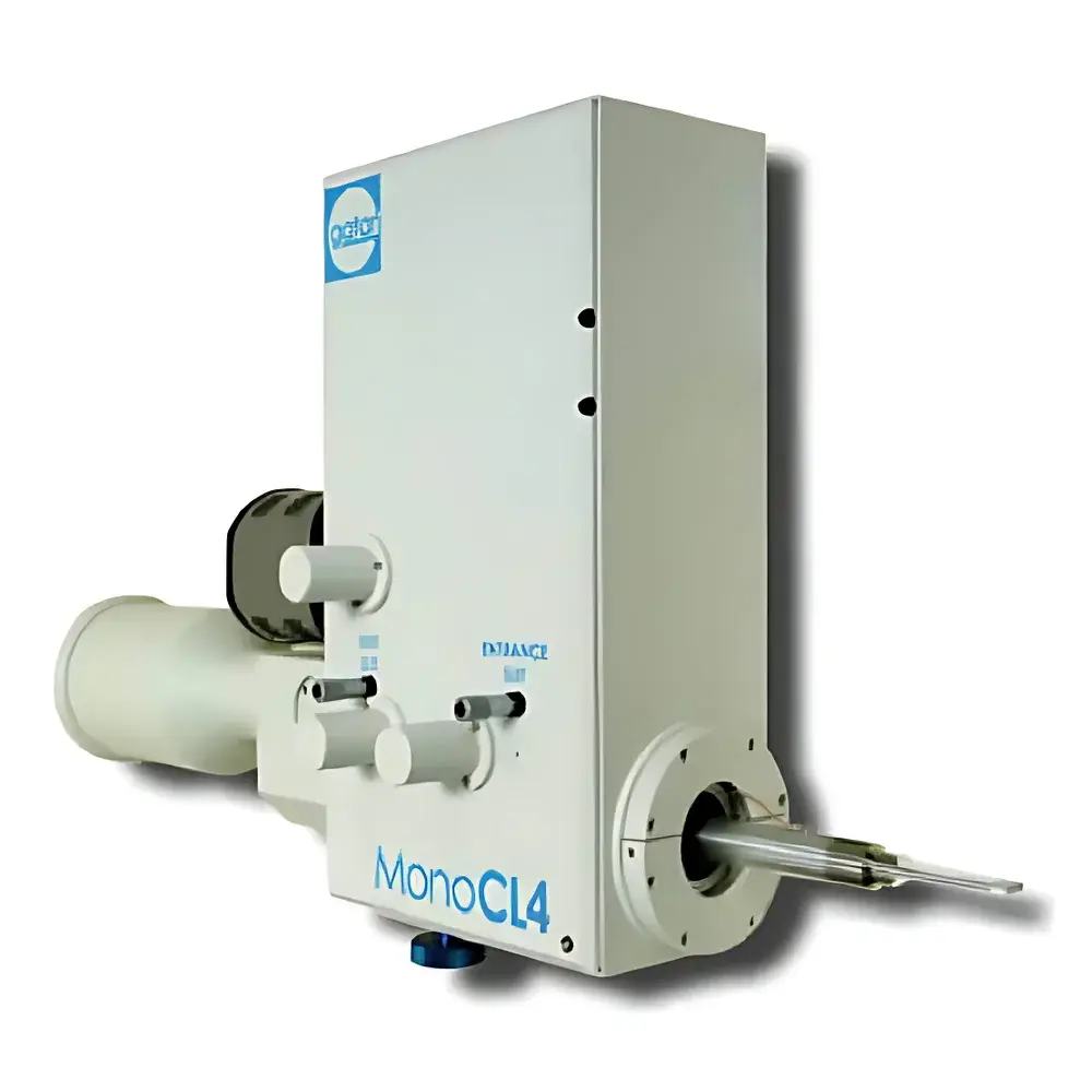
Gatan MonoCL4 High-Resolution Cathodoluminescence Imaging and Spectroscopy System
| Brand | Gatan |
|---|---|
| Origin | USA |
| Model | MonoCL4 |
| Manufacturer | Gatan, Inc. |
| Detector Type | Parabolic Mirror Collection with Direct-Coupled Czerny-Turner Monochromator |
| Spectral Range | 185–850 nm |
| Spectral Resolution | Adjustable via Micrometer Slit (Typical FWHM < 1.5 nm at 500 nm) |
| Collection Efficiency | Optimized via Diamond-Turned Parabolic Mirror (75 mm retractable stroke) |
| Detection | PMT with Integrated Preamplifier |
| Software | DigitalMicrograph® with MonoCL4 Plugin |
| Compliance | Fully Compatible with SEM/TEM/EPMA Platforms |
Overview
The Gatan MonoCL4 is a high-performance cathodoluminescence (CL) imaging and spectroscopy system engineered for integration with scanning electron microscopes (SEM), transmission electron microscopes (TEM), and electron probe microanalyzers (EPMA). It operates on the fundamental principle of electron-beam-induced photon emission from inelastic scattering events within solid-state materials. When a focused electron beam excites a specimen, bound electrons are promoted to higher energy states; subsequent radiative recombination produces photons whose wavelength, intensity, and spatial distribution encode critical information about electronic band structure, defect states, compositional heterogeneity, strain fields, and crystallinity. The MonoCL4 captures this signal with exceptional optical throughput and spectral fidelity—leveraging a diamond-turned parabolic collection mirror directly coupled to a high-efficiency Czerny-Turner monochromator and photon-counting photomultiplier tube (PMT). This architecture minimizes optical losses across the deep-UV to near-IR range (185–850 nm), enabling quantitative CL mapping at nanoscale spatial resolution without inducing beam damage or non-equilibrium artifacts.
Key Features
- Optimized optical path: Retractable, detachable parabolic mirror (75 mm stroke, 8.75 mm thickness) with integrated LED position indicator ensures repeatable alignment and maximal solid-angle collection.
- Direct-coupled cavity monochromator: Eliminates intermediate optics, preserving étendue and signal-to-noise ratio across the full spectral range.
- Motorized mode switching: Seamless transition between panchromatic imaging and monochromatic acquisition via motor-driven dichroic mirror.
- Tunable spectral resolution: Precision micrometer slit (0.01 mm increments) allows adjustment of bandwidth from 10 nm FWHM, supporting both high-resolution spectroscopy and rapid hyperspectral mapping.
- Calibrated spectral response: Factory-measured system response curves (350–850 nm) and built-in ITS-L calibration lamp enable traceable intensity quantification and inter-instrument comparability.
- Digital control architecture: PA4 controller synchronizes monochromator positioning, PMT gain, and detector timing with sub-millisecond precision; remote manual controller provides real-time HV readout and imaging parameter adjustment.
- Integrated software suite: DigitalMicrograph® platform with dedicated MonoCL4 plugin enables automated spectral acquisition, Gaussian deconvolution of multi-peak spectra, batch processing of CL maps, and export to standard scientific formats (e.g., TIFF, HDF5, CSV).
Sample Compatibility & Compliance
The MonoCL4 supports diverse specimen geometries—including bulk minerals, polished thin sections, TEM lamellae, nanowires, quantum dots, and epitaxial semiconductor heterostructures—without requiring conductive coatings in most cases. Its low-dose operation (<100 pA beam current) preserves beam-sensitive phases while maintaining sufficient signal for spectral decomposition. The system complies with international standards for analytical electron microscopy workflows: spectral data adhere to ASTM E1508 (Standard Guide for Quantitative Analysis by Energy-Dispersive Spectroscopy) metadata conventions; acquisition logs include timestamped instrument parameters, detector settings, and stage coordinates—enabling full traceability per ISO/IEC 17025 and FDA 21 CFR Part 11 requirements when deployed in regulated environments. All hardware interfaces conform to SEM manufacturer protocols (e.g., Zeiss, Thermo Fisher, JEOL), ensuring stable synchronization with beam scanning, stage motion, and detector gating.
Software & Data Management
DigitalMicrograph® serves as the unified control and analysis environment. Upon startup, the MonoCL4 plugin executes an automated spectral calibration routine using the internal ITS-L lamp, correcting for grating efficiency drift and detector sensitivity variations. Spectral acquisition supports single-point, line-scan, and raster-based hyperspectral modes—with real-time preview of intensity-weighted RGB composites and spectral libraries. Post-acquisition tools include peak fitting (multi-Gaussian, Lorentzian, Voigt), background subtraction (polynomial, SNIP), spatial correlation overlays with EDS/BSE/EBSD datasets, and batch normalization across multi-session experiments. Data files embed EXIF-like metadata (beam kV, working distance, dwell time, slit width, grating order) and support DICOM-SR export for cross-platform archival. Scripting via DM’s Python API allows custom automation of QA/QC routines, report generation, and integration with LIMS systems.
Applications
In geosciences, the MonoCL4 resolves growth zoning in zircon, identifies fluid inclusion healing textures in quartz, and discriminates Mn²⁺- vs. Fe³⁺-activated luminescence in feldspars—providing petrogenetic constraints inaccessible via conventional BSE or EDS. In semiconductor research, it maps threading dislocation densities in GaN epilayers, correlates carrier localization with InGaN quantum well composition gradients, and quantifies oxygen vacancy signatures in transparent conducting oxides (e.g., ZnO, ITO). For photovoltaics, its UV sensitivity enables direct probing of silicon defect-related luminescence (D-lines, W-lines) despite Si’s weak CL yield. In forensics and pharmaceuticals, it delivers spectral fingerprints of pigment particles, counterfeit drug excipients, and biomineralized tissues—complementing fluorescence and Raman modalities with superior spatial resolution.
FAQ
What vacuum compatibility does the MonoCL4 require?
The system is rated for ultra-high vacuum (UHV) conditions down to 1×10⁻⁷ Pa and integrates seamlessly with standard SEM/TEM column vacuum architectures via ConFlat flanges.
Can the MonoCL4 be retrofitted to older SEM models?
Yes—compatible with all major SEM platforms manufactured since 2005, provided mechanical mounting space and electrical interface access (RS-232, USB, or Ethernet) are available.
Is spectral calibration traceable to NIST standards?
While the ITS-L lamp provides internal reproducibility, users may perform external calibration using NIST-traceable emission standards (e.g., Hg/Ar lamps) via the system’s open optical port.
How is data integrity ensured during long-duration mapping sessions?
DigitalMicrograph implements checksummed HDF5 storage with embedded acquisition metadata, automatic backup on controller failure, and optional encrypted write-to-disk policies compliant with ISO 27001 frameworks.
Does the system support time-resolved CL measurements?
Not natively—however, external beam blanking signals can be synchronized with the PA4 controller for pulsed-beam experiments, enabling lifetime estimation via gated detection when paired with fast-scanning SEMs.


