HeadScan Human Imaging Photographic Studio
| Brand | ORION |
|---|---|
| Origin | France |
| Manufacturer Type | Authorized Distributor |
| Product Origin | Imported |
| Model | HeadScan |
| Pricing | Available Upon Request |
Overview
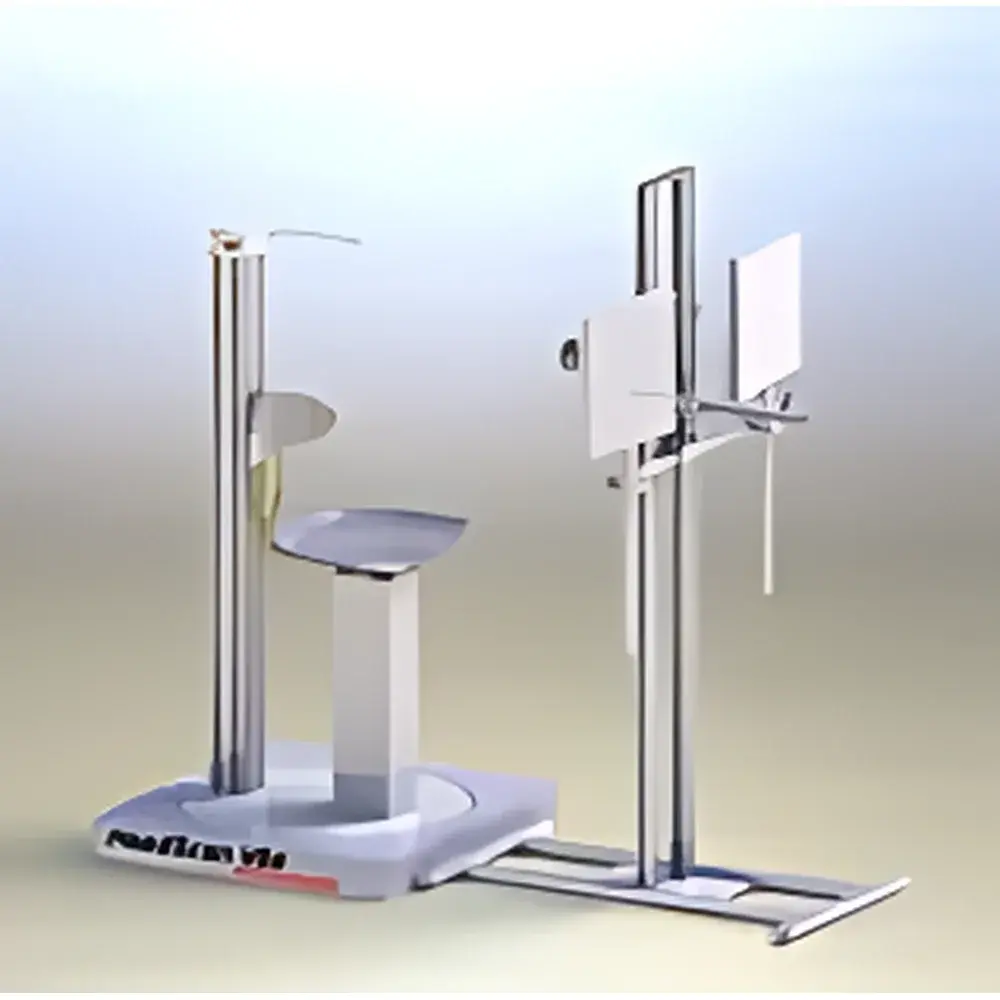
The HeadScan Human Imaging Photographic Studio is a calibrated, standardized imaging environment engineered for high-fidelity acquisition of facial and full-body surface images under controlled illumination, geometry, and spectral conditions. Designed specifically for dermatological research and cosmetic efficacy evaluation, the system operates on the principles of quantitative digital photogrammetry and multispectral reflectance imaging. It enables objective, repeatable documentation of skin surface morphology, chromatic distribution, texture topography, and geometric contour—critical for longitudinal clinical trials, regulatory submissions (e.g., ISO 16128, COLIPA guidelines), and comparative product performance studies. Unlike generic photography setups, HeadScan integrates fixed-position lighting arrays (D65-standard illuminants), motorized subject positioning, and synchronized camera triggering to eliminate operator-induced variability in image acquisition.
Key Features
- Modular studio architecture with adjustable backdrop systems (neutral gray and spectrally neutral white) compliant with ISO 20772:2018 for skin imaging standardization.
- Integrated multi-angle illumination rig featuring tunable D65 daylight simulation (CCT 6500 K ± 100 K, CRI >95) with shadow-free uniformity across target zones (±3% irradiance variation over 30 × 40 cm field).
- Motorized, height- and tilt-adjustable subject chair with anthropometric reference markers (Frankfurt plane alignment indicators) for reproducible head and torso positioning.
- High-resolution industrial-grade CMOS sensor (≥24 MP, 12-bit dynamic range) with macro lens capability (1:1 magnification at 30 cm working distance) for localized lesion or wrinkle analysis.
- Hardware-synchronized dual-camera configuration (frontal + oblique 45°) enabling stereoscopic surface reconstruction and depth-aware morphometric quantification.
- Calibration-certified color targets (X-Rite ColorChecker Passport) and spatial scale bars embedded in each image frame for traceable photometric and geometric metrology.
Sample Compatibility & Compliance
The HeadScan system accommodates adult and adolescent human subjects across diverse Fitzpatrick skin types (I–VI). It supports static pose acquisition for face, neck, décolletage, hands, thighs, abdomen, and back regions. All imaging protocols adhere to ISO/IEC 17025-accredited laboratory practices when operated within GLP-compliant environments. The system is validated for use in studies referenced by EU Cosmetics Regulation (EC No. 1223/2009), US FDA guidance on cosmetic labeling claims, and ICH E6(R3) clinical trial standards. Image metadata includes EXIF tags compliant with DICOM Supplement 171 (Dermatology Imaging) for interoperability with PACS and clinical trial databases.
Software & Data Management
FrameScan software—bundled with HeadScan—is a CE-marked Class I medical device application (MDD 93/42/EEC) for quantitative dermal image analysis. It provides FDA 21 CFR Part 11-compliant audit trails, user role-based access control, and encrypted local storage. Core analytical modules include: (i) CIELAB-based chroma mapping for melanin/erythema index derivation (ΔL*, Δa*, Δb*); (ii) grayscale gradient thresholding and Hessian-based ridge detection for wrinkle depth/length quantification; (iii) ellipse-fitting and contour segmentation algorithms for spot area/perimeter/aspect ratio measurement; (iv) 3D surface mesh generation from stereo pairs for curvature and volume change assessment (e.g., post-treatment edema or contouring effects). Raw image exports support TIFF (uncompressed), JPEG2000 (lossless), and DICOM-SR formats.
Applications
- Objective evaluation of skin brightening agents via longitudinal L* (lightness) and b* (yellowness) tracking across standardized ROI masks.
- Stability assessment of lipsticks and foundations through time-series color drift analysis (ΔE00) under accelerated aging conditions (UV exposure, temperature cycling).
- Quantitative characterization of hyperpigmented macules—including area, density, and spatial clustering metrics—aligned with PIH (post-inflammatory hyperpigmentation) scoring criteria.
- Morphometric profiling of facial aging: nasolabial fold depth, periorbital wrinkle count, and mandibular angle definition using landmark-free deep learning-assisted segmentation.
- Body contour analysis for aesthetic interventions: pre-/post-procedure volumetric comparison of abdominal wall convexity or thigh circumference profiles using geodesic surface distance mapping.
FAQ
Is HeadScan compatible with third-party image analysis platforms?
Yes—FrameScan exports annotated datasets in open formats (TIFF, CSV, DICOM-SR) and supports API-driven integration with MATLAB, Python (OpenCV, scikit-image), and commercial platforms including Visage Imaging and DermEngine.
Does the system require annual recalibration?
ORION recommends biannual verification of illuminant spectral output and camera gamma response using NIST-traceable calibration kits; full system validation is advised prior to pivotal clinical study initiation.
Can HeadScan be deployed in mobile clinical trial units?
The modular design supports rapid reassembly in ISO Class 8 cleanroom trailers or GMP-aligned mobile labs; power requirements are 230 V AC ±10%, 50 Hz, with optional UPS integration.
What regulatory documentation is provided with delivery?
Each unit ships with EU Declaration of Conformity, ISO 13485 manufacturing certificate (applies to FrameScan software), calibration certificates for lighting and optics, and a comprehensive SOP suite aligned with ISO/IEC 17025 clause 7.8.