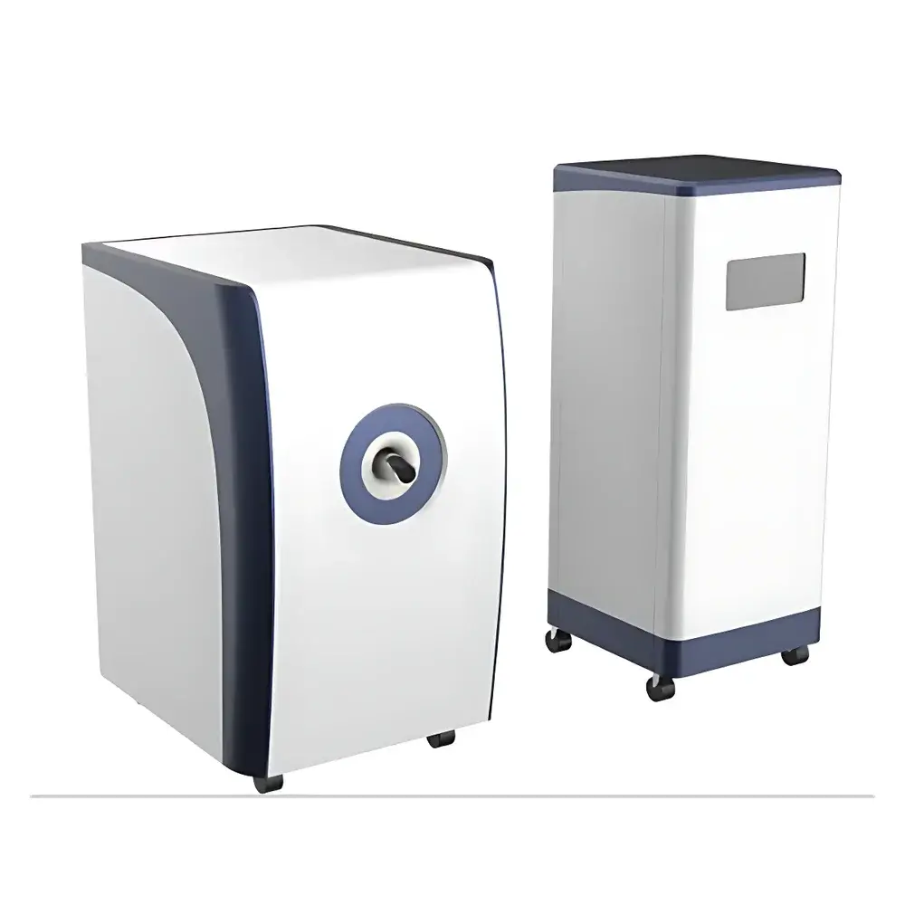
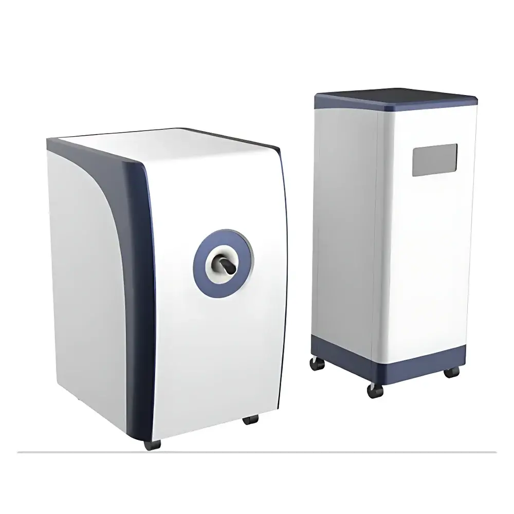
HTNMR HT-MRSI60-25 1.5T Small Animal In Vivo Magnetic Resonance Imaging and Spectroscopic Imaging System
| Brand | HTNMR |
|---|---|
| Origin | Shanghai, China |
| Manufacturer Type | Original Equipment Manufacturer (OEM) |
| Product Category | Domestic |
| Model | HT-MRSI60-25 |
| Instrument Type | Magnetic Resonance Imaging (MRI) & Magnetic Resonance Spectroscopic Imaging (MRSI) System |
| Spatial Resolution | 0.8 ppm (chemical shift resolution) |
| Field of View (FOV) | 25 mm × 30 mm |
| Magnetic Field Strength | 1.5 T |
| ¹H Resonance Frequency | 55–59 MHz |
| Homogeneity | <2 ppm over 25 mm DSV |
| Temperature Stability | ±0.06 K/h after 2-h warm-up |
| SNR | 55 dB |
| Geometric Distortion | <1% |
| Image Matrix | Up to 512 × 512 (2D), 128 × 128 × 128 (3D), 64 × 64 × 64 × 64 (4D MRSI) |
Overview
The HTNMR HT-MRSI60-25 is a preclinical 1.5 Tesla magnetic resonance imaging and spectroscopic imaging (MRSI) system engineered for high-fidelity in vivo structural, functional, and metabolic characterization of small animal models—including mice, rats, and small plant or invertebrate specimens. Operating on the physical principles of nuclear magnetic resonance (NMR), this system detects radiofrequency signals emitted by hydrogen (¹H) nuclei under controlled static and gradient magnetic fields. Unlike conventional MRI systems that emphasize anatomical contrast, the HT-MRSI60-25 integrates spatial encoding with chemical shift dispersion—enabling simultaneous acquisition of 3D anatomical images and 1D spectral data across each voxel. This four-dimensional (4D) capability—three spatial dimensions plus one spectral dimension—supports non-invasive, label-free molecular phenotyping in longitudinal studies. The system employs a high-homogeneity permanent magnet architecture with deuterium-based field-frequency lock (D₂O external lock), ensuring field stability better than 10 Hz/month and operational robustness across ambient temperatures from 10 °C to 35 °C without active shielding or cryogen dependency.
Key Features
- Integrated 1.5 T permanent magnet with <2 ppm field homogeneity over a 25 mm diameter spherical volume (DSV)
- Dual-mode acquisition: high-resolution 3D/4D MRI and MRSI using customizable pulse sequences
- Comprehensive pulse sequence library including Spin Echo (SE), Gradient Echo (GE), Echo Planar Imaging (EPI), Inversion Recovery (IR), Dixon water-fat separation, and Echo Time Encoding (ETE) spectroscopic imaging
- Four-dimensional Fourier-transform SE acquisition (64 × 64 × 64 × 64 matrix support) for volumetric metabolite mapping
- Dedicated workstation with real-time reconstruction engine for large-volume k-space and spectral data processing
- Modular software platform supporting pulse sequence editing, relaxation time quantification (T₁/T₂ mapping), and 3D surface rendering with pseudo-color spectral overlays
- Active temperature stabilization (±0.06 K/h drift post-warm-up) enabling reproducible multi-session acquisitions
Sample Compatibility & Compliance
The HT-MRSI60-25 accommodates live small animal subjects up to 25 mm in diameter for 3D imaging and 20 mm for full 4D MRSI acquisition. Its RF coil design supports both transmit/receive quadrature detection and optional phased-array configurations for improved SNR in targeted regions. The system complies with IEC 61000-6-3 (EMC emission standards) and meets essential safety requirements outlined in ISO 10974 for MR equipment used in biomedical research. While not intended for clinical diagnostic use, all imaging protocols align with preclinical best practices defined by the NIH Office of Laboratory Animal Welfare (OLAW) and AAALAC International guidelines. Data acquisition workflows support audit-ready metadata logging—including pulse sequence parameters, gradient timing, RF amplitude calibration, and environmental monitoring—facilitating GLP-compliant study documentation.
Software & Data Management
The proprietary acquisition and analysis suite provides native support for DICOM 3.0 export, NIfTI-1 format conversion, and Bruker ParaVision-compatible parameter files. Pulse sequence programming is implemented via a Python-based scripting interface compliant with ISMRM Pulse Sequence Description Language (PSDL) conventions. All raw k-space and spectral datasets are stored with embedded acquisition metadata (e.g., TR/TE/TI, bandwidth, voxel size, shimming history). The reconstruction module implements parallel imaging acceleration (SENSE-equivalent), partial Fourier reconstruction, and spectral apodization with exponential line-broadening or Gaussian filtering. Quantitative analysis tools include voxel-wise T₂ mapping using mono-exponential fitting, Dixon-based fat fraction calculation, and spectral peak integration using Lorentzian/Gaussian deconvolution. Audit trails record user actions, parameter modifications, and software versioning per FDA 21 CFR Part 11 Annex 11 recommendations for regulated research environments.
Applications
This system serves as a core platform for translational neuroscience, oncology, and metabolic disease research. Typical use cases include longitudinal tracking of neurodegenerative biomarkers in Alzheimer’s mouse models via hippocampal MRSI; in vivo assessment of tumor metabolism in orthotopic glioblastoma xenografts using choline/NAA/Cr ratio quantification; evaluation of myocardial fat infiltration in diet-induced obesity models via Dixon imaging; and developmental studies in zebrafish or Arabidopsis thaliana using low-SAR GE-EPI for motion-robust functional imaging. Its open-architecture design also enables method development in advanced techniques such as chemical exchange saturation transfer (CEST), diffusion tensor imaging (DTI), and dynamic contrast-enhanced (DCE) pharmacokinetic modeling—subject to appropriate RF coil and gradient performance validation.
FAQ
What is the difference between MRI and MRSI on this system?
MRSI extends standard MRI by adding spectral encoding along a fourth dimension—allowing spatial localization of metabolite-specific resonance frequencies (e.g., lactate at 1.33 ppm, NAA at 2.02 ppm) within each image voxel.
Can the system perform quantitative T₁ and T₂ mapping?
Yes—the included relaxation time acquisition module supports variable TR/TE sampling and nonlinear least-squares fitting to generate parametric T₁ and T₂ maps with pixel-wise uncertainty estimation.
Is the system compatible with third-party analysis software like FSL or SPM?
Raw data exports in NIfTI-1 and DICOM formats ensure full interoperability with major neuroimaging toolkits, including FSL, SPM, AFNI, and MRtrix3.
Does the system require liquid helium or other cryogens?
No—it utilizes a passively shielded permanent magnet with D₂O field-frequency lock, eliminating cryogenic infrastructure and associated maintenance costs.
What regulatory documentation is provided for institutional biosafety or ethics review?
HTNMR supplies a comprehensive technical dossier including magnetic field maps, SAR calculations per IEC 62704, RF exposure reports, and compliance statements aligned with ISO/IEC 17025-accredited test protocols.