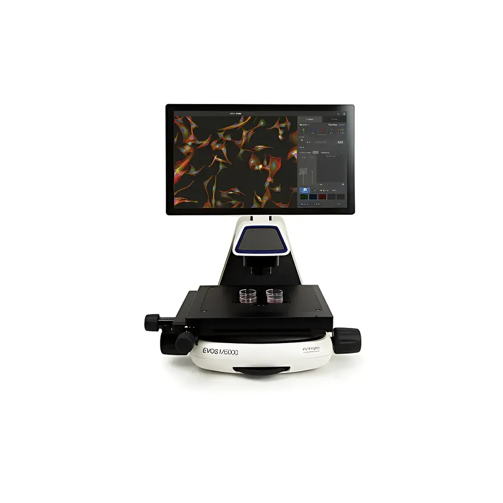
Invitrogen EVOS M5000 Intelligent Fluorescence Cell Imaging System
| Brand | Invitrogen |
|---|---|
| Origin | USA |
| Manufacturer Type | Original Equipment Manufacturer (OEM) |
| Import Status | Imported |
| Model | EVOS M5000 |
| Pricing | Available Upon Request |
Overview
The Invitrogen EVOS M5000 Intelligent Fluorescence Cell Imaging System is a fully integrated, upright-style automated microscope platform engineered for quantitative live and fixed-cell imaging in academic, pharmaceutical, and biotechnology laboratories. Built upon a precision optical architecture combining high-quantum-efficiency monochrome CMOS imaging with proprietary multi-spectral LED illumination, the system enables simultaneous acquisition of fluorescence and true-color brightfield images without mechanical filter wheel movement or lamp alignment. Its core optical design follows Köhler illumination principles with optimized light path geometry to minimize chromatic aberration and maximize signal-to-noise ratio across excitation/emission bands. The system supports objective-based magnification ranging from 2× to 60×, with motorized nosepiece and autofocus-driven Z-axis control enabling reproducible, drift-compensated image capture—critical for longitudinal studies and regulatory-compliant workflows.
Key Features
- High-sensitivity monochrome CMOS sensor (≥4.2 MP resolution) with >75% quantum efficiency at 520 nm, optimized for low-light fluorescence detection including GFP, RFP, Cy3, and DAPI.
- Dual-mode illumination engine: Independently controllable LED arrays for fluorescence excitation (365–630 nm range) and color-accurate brightfield illumination (CRI >90), enabling faithful H&E and IHC image reproduction without post-acquisition color correction.
- Motorized Z-stack acquisition with sub-micron step resolution (down to 0.1 µm), supporting maximum intensity projection, extended depth-of-field synthesis, and volumetric reconstruction of thick specimens up to 200 µm.
- Integrated autofocus algorithm utilizing contrast-based and pattern-recognition methods; maintains focus stability over ≥24-hour time-lapse sessions with thermal drift compensation.
- Modular environmental control compatibility: Designed to interface with the EVOS Onstage Incubator for precise regulation of temperature (±0.2°C), CO₂ (0–20%), O₂ (0.1–21%), and relative humidity (up to 95% RH) during long-term live-cell imaging.
- Touch-optimized GUI with one-click multi-channel acquisition, real-time channel blending, and on-the-fly exposure/gain adjustment—no scripting or third-party software required.
Sample Compatibility & Compliance
The EVOS M5000 accommodates standard microscopy formats including glass-bottom dishes (35 mm, 60 mm), chamber slides (1–8 wells), and histological sections on 1–2 mm thick glass slides. It supports common fluorescent probes (e.g., Image-iT Hypoxia Reagents, NucBlue Live, ReadyProbes viability dyes) and immunolabeling protocols validated under ISO 13485-aligned manufacturing processes. Data acquisition and metadata logging comply with ALP/CLIA-relevant documentation standards; audit trail functionality meets GLP and FDA 21 CFR Part 11 requirements when used with optional secure user authentication and electronic signature modules. All optical components are certified per ISO 10110-7 for surface quality and wavefront error.
Software & Data Management
The embedded EVOS Imaging Software v3.x provides native support for TIFF, OME-TIFF, and JPEG2000 export with embedded EXIF-style metadata (objective ID, exposure time, LED intensity, Z-position, timestamp). Batch processing includes auto-thresholding, object segmentation via trainable pixel classifiers, and morphometric analysis (cell count, area, circularity, intensity distribution). Exported datasets are compatible with FIJI/ImageJ, MATLAB, and Python-based analysis pipelines (e.g., scikit-image, napari). Raw image data integrity is preserved through lossless compression and SHA-256 checksum generation per acquisition session. Local storage utilizes encrypted SSD with configurable auto-archive to network-attached storage (NAS) or LIMS-integrated repositories.
Applications
- Quantitative hypoxia profiling using ratiometric O₂-sensitive dyes in cancer spheroids and organoids.
- Time-lapse tracking of neuronal outgrowth, dendritic spine dynamics, and synaptic vesicle trafficking under controlled gas environments.
- Automated viability assessment in primary T-cell expansion assays using dual nuclear staining (DAPI/NucGreen Dead 488) with machine-learning-assisted dead-cell classification.
- 3D reconstruction of vascularized tissue constructs and scaffold-based co-cultures imaged across ≥15 Z-planes.
- Standardized IHC scoring for translational pathology studies requiring color-fidelity preservation across serial sections.
- Preclinical assay development compliant with ASTM E3152-18 (standard guide for digital pathology image acquisition).
FAQ
Does the EVOS M5000 support oil immersion objectives?
Yes—motorized nosepiece accepts standard RMS-threaded 60× and 100× oil-immersion objectives with integrated coverslip correction collars.
Can acquired Z-stacks be exported as 3D volume files for external rendering?
Yes—OME-TIFF stacks include dimensional metadata and can be imported directly into Imaris, Arivis Vision4D, or Blender for surface rendering and quantitative volumetric analysis.
Is remote operation supported for shared core facility deployment?
The system supports VNC-based remote desktop access and RESTful API endpoints for instrument control and metadata retrieval via institutional authentication servers.
What calibration standards are provided for fluorescence intensity quantification?
NIST-traceable fluorescent microsphere reference kits (e.g., Chroma 40200 series) are recommended for daily photometric calibration; system linearity is verified per ISO 21530 across 4-log dynamic range.
How is focus drift managed during extended incubator-based time-lapse experiments?
Real-time focus feedback is derived from continuous low-intensity IR LED illumination and dedicated focus sensor module, correcting positional drift at ≤10-second intervals without perturbing sample physiology.


