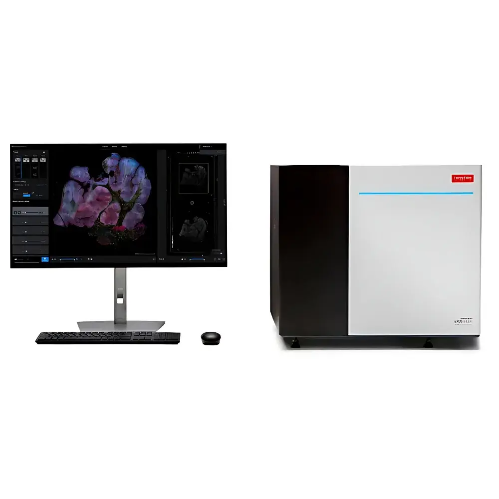
Invitrogen™ EVOS™ S1000 Spatial Biology Imaging Analysis System
| Brand | Thermo Fisher Scientific |
|---|---|
| Origin | Shanghai, China |
| Manufacturer Type | Authorized Distributor |
| Origin Category | Domestic (China-manufactured under Thermo Fisher specifications) |
| Model | Invitrogen™ EVOS™ S1000 |
| Pricing | Available upon Request |
Overview
The Invitrogen™ EVOS™ S1000 Spatial Biology Imaging Analysis System is a benchtop, fully integrated digital microscope platform engineered for high-content, multiplexed spatial phenotyping of intact tissue sections. It employs widefield fluorescence imaging combined with spectral unmixing technology to resolve overlapping emission spectra from up to nine co-localized biomarkers in a single acquisition—without requiring sequential staining or wash cycles. Designed specifically for translational research and preclinical development workflows, the system bridges the gap between conventional histopathology and next-generation spatial omics by delivering quantitative, location-resolved protein and nucleic acid expression data directly from formalin-fixed paraffin-embedded (FFPE) or frozen tissue specimens. Its optical architecture supports simultaneous multispectral capture across the visible and near-visible range (400–720 nm), enabling robust signal separation even with conventional fluorophores exhibiting significant spectral overlap.
Key Features
- Multiplex Imaging Efficiency: Acquires full-spectrum, 9-plex images over a 1 cm² tissue area in ≤20 minutes at 10× magnification—including automated stage scanning, spectral acquisition, pixel-level unmixing, and seamless tile stitching.
- Modular Optical Configuration: Equipped with a motorized 5-position objective turret supporting 2.5×, 10×, 20×, 40×, and optional 60× plan-apochromat objectives; all optimized for high numerical aperture and uniform flat-field illumination.
- Scientific-Grade Detection: Integrated 4.2-megapixel, 16-bit sCMOS sensor with >82% quantum efficiency at 550 nm and sub-electron read noise—enabling detection of low-abundance targets in heterogeneous tissue microenvironments.
- Spectral Unmixing Engine: Proprietary real-time linear unmixing algorithm trained on reference spectra of common fluorophores (e.g., Alexa Fluor 488, 555, 647; Cy3, Cy5; DAPI), minimizing cross-talk and preserving spatial fidelity without iterative deconvolution.
- Intelligent Autofocus: Dual-mode autofocus system combining laser-based Z-height mapping and contrast-based software-assisted refinement—ensuring consistent focus across variable tissue thicknesses and section folds.
- Workflow-Centric Software: EVOS Imaging Software v3.x provides guided acquisition protocols, region-of-interest (ROI) annotation, batch processing, and export to OME-TIFF format with embedded metadata compliant with Bio-Formats and QuPath interoperability standards.
Sample Compatibility & Compliance
The EVOS™ S1000 accepts standard glass slides (1 × 3 inches / 25 × 75 mm), including charged, silanized, and polymer-coated variants. It supports immunofluorescence (IF), immunohistochemistry (IHC), RNAscope®-based in situ hybridization (ISH), multiplexed IF (mIF), and chromogenic assays on FFPE and cryosections up to 20 µm thick. All hardware and firmware comply with IEC 61010-1:2012 safety requirements for laboratory equipment. Data handling adheres to ALCOA+ principles (Attributable, Legible, Contemporaneous, Original, Accurate, Complete, Consistent, Enduring, Available); audit trail functionality meets FDA 21 CFR Part 11 readiness when deployed in validated environments under GLP or GMP-aligned SOPs.
Software & Data Management
EVOS Imaging Software delivers a role-based interface with three user profiles: Operator (acquisition-only), Analyst (quantification + annotation), and Administrator (system configuration + audit log review). Image data are stored locally in hierarchical folder structures with SHA-256 checksum validation. Export options include multi-channel TIFF stacks, OME-TIFF with structured metadata (including objective magnification, exposure time, gain, and spectral band assignment), and CSV-based cell-level feature tables compatible with downstream analysis in CellProfiler, HALO®, Visium Spatial Gene Expression pipelines, and custom Python/R scripts. The software supports DICOM-SR export for integration into PACS-enabled pathology informatics systems.
Applications
- Translational biomarker discovery in oncology, neurology, and immunology tissue cohorts
- Validation of spatial transcriptomics findings via orthogonal protein-level mapping
- Quantitative assessment of tumor immune microenvironment (TIME) architecture—e.g., PD-L1⁺/CD8⁺ proximity metrics, tertiary lymphoid structure (TLS) quantification
- Preclinical toxicology studies requiring spatial resolution of drug-induced morphological and molecular changes
- Quality control of tissue microarrays (TMAs) and digital pathology slide digitization workflows
- Academic core facility deployment for shared-resource multiplex imaging services
FAQ
Is the EVOS™ S1000 suitable for live-cell imaging?
No—it is optimized for fixed-tissue analysis only. The system lacks environmental control (temperature, CO₂, humidity) and high-speed acquisition modes required for dynamic cellular processes.
Can third-party antibodies or probes be used with this system?
Yes. The platform imposes no proprietary reagent restrictions; compatibility depends solely on spectral characteristics and recommended labeling protocols for the selected fluorophores.
Does the system support whole-slide scanning at 40×?
Yes—automated mosaic acquisition is available at all supported magnifications, with adaptive focus correction applied per tile to accommodate tissue topography.
What level of IT infrastructure is required for data storage and analysis?
A minimum of 16 GB RAM, 1 TB SSD, and Windows 10/11 Pro (64-bit) is recommended. For large-scale cohort studies, network-attached storage (NAS) integration and GPU-accelerated unmixing plugins are supported via optional software modules.
Is remote operation or multi-user access supported?
Local network sharing of acquisition queues and image repositories is enabled; however, concurrent remote control requires external VDI or terminal server configuration—not natively embedded in the base software stack.
Related Products
")


