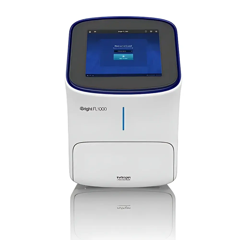
Invitrogen iBright FL1000 Smart Gel Imaging System
| Brand | Invitrogen |
|---|---|
| Origin | Singapore |
| Manufacturer Type | Original Equipment Manufacturer (OEM) |
| Product Category | Imported Instrument |
| Model | FL1000 |
| Instrument Type | Multicolor Fluorescent Gel Imaging System |
| CCD Resolution | 9.1 MP |
| Bit Depth | 16-bit |
| CCD Sensor Size | 1-inch |
| Lens | 25 mm f/0.95 Fixed Focus |
| Light Source | Green LED (for nucleic acid staining), Optional NIR/RGB Excitation Modules |
| Display | 12-inch Multi-Touch LCD |
| Imaging Area | Supports up to 4 mini-gels or 2 mid-size gels simultaneously |
| Software Platform | iBright Analysis Software on Thermo Fisher Cloud |
| Compliance | Designed for GLP/GMP-adjacent workflows |
Overview
The Invitrogen iBright FL1000 Smart Gel Imaging System is a fully integrated, cloud-connected platform engineered for quantitative and qualitative analysis of nucleic acids, proteins, and post-translational modifications across multiple detection modalities. Utilizing a thermoelectrically cooled scientific-grade CCD sensor with 9.1 megapixel resolution and true 16-bit digitization, the FL1000 captures high-fidelity images with exceptional dynamic range and low-noise performance—critical for reproducible chemiluminescent Western blot quantification and multiplex fluorescent detection. Unlike legacy UV transilluminators, the system employs a dedicated green LED excitation source (λ ≈ 530 nm) optimized for SYBR Safe, GelGreen, and other visible-light–compatible dyes—eliminating UV-associated DNA damage and operator exposure risks while maintaining high sensitivity for agarose and polyacrylamide gel imaging. Its optical architecture centers on a large-aperture f/0.95 fixed-focus lens, enabling maximum photon collection efficiency and consistent depth-of-field across the entire 1-inch sensor area.
Key Features
- Thermoelectrically cooled CCD detector with 9.1 MP resolution and 16-bit analog-to-digital conversion for precise grayscale linearity and wide signal capture range.
- Dedicated green LED transillumination (530 nm) compliant with IEC 62471 photobiological safety standards—replacing hazardous short-wave UV sources without compromising stain sensitivity.
- Integrated multi-channel fluorescence capability supporting simultaneous detection in RGB and near-infrared (NIR) channels—enabling 4-color multiplex Western blotting using IRDye and Alexa Fluor conjugates.
- 12-inch capacitive multi-touch display with gesture-based navigation (pinch-to-zoom, drag-to-pan, double-tap focus)—designed for glove-compatible operation in biosafety cabinets and core facility environments.
- Smart Explore automated imaging protocol: performs real-time exposure optimization, auto-focus calibration, sample alignment, and background subtraction—all executed in under 30 seconds per acquisition.
- Large-format imaging chamber accommodates up to four 8 × 10 cm mini-gels or two 13.5 × 10 cm mid-size gels in a single acquisition—reducing inter-run variability and increasing throughput.
Sample Compatibility & Compliance
The iBright FL1000 supports standard electrophoretic formats including SDS-PAGE, native PAGE, 2D gels, agarose gels, and capillary electrophoresis strips. It is validated for use with common stains and probes: Coomassie Brilliant Blue R-250, Silver Nitrate, SYPRO Ruby, ethidium bromide alternatives (e.g., GelRed, SYBR Gold), and chemiluminescent substrates (e.g., Luminata Forte, SuperSignal West Femto). While not certified as a medical device, its software architecture aligns with principles outlined in FDA 21 CFR Part 11 for electronic records and signatures—supporting user-defined roles, password-protected access, and immutable audit trails for image metadata, acquisition parameters, and analysis history. Instrument usage logs, firmware version tracking, and remote diagnostics are accessible via Thermo Fisher Connect—a secure, HIPAA-aligned infrastructure used by academic, clinical, and contract research organizations.
Software & Data Management
iBright Analysis Software operates natively within the Thermo Fisher Cloud environment, requiring no local installation or license server. All image acquisition, annotation, band intensity quantification (with background correction and molecular weight calibration), and publication-ready figure generation occur through a browser-based interface compatible with Chrome, Edge, and Safari. Raw .TIFF files retain full 16-bit depth and embedded EXIF metadata—including exposure time, gain setting, lens aperture, and temperature-stabilized sensor status. Export options include PDF reports with embedded traceable metadata, CSV-formatted intensity tables for statistical analysis in R or Python, and FAIR-compliant JSON-LD manifests. Data retention policies, sharing permissions (view/edit/collaborate), and version-controlled project folders are centrally managed by institutional IT administrators.
Applications
- Quantitative Western blot analysis using enhanced chemiluminescence (ECL) and near-infrared fluorescence (NIRF) detection—ideal for phosphoprotein profiling and low-abundance target validation.
- Multiplex nucleic acid detection in qPCR product verification, CRISPR editing efficiency assessment, and methylation-specific PCR (MSP) assays.
- High-throughput screening of protein expression libraries, antibody specificity testing, and quality control of recombinant therapeutics.
- Educational laboratory instruction—leveraging intuitive touch interface and guided workflows to reduce training time for undergraduate and graduate students.
- Core facility deployment—where centralized instrument monitoring, usage billing integration, and cross-platform data interoperability (via RESTful API endpoints) streamline resource allocation.
FAQ
Does the iBright FL1000 support time-lapse chemiluminescent imaging?
Yes—the system allows programmable sequential acquisitions with variable exposure durations and automatic gain adjustment, enabling kinetic monitoring of ECL signal decay for optimal integration window selection.
Can third-party analysis tools import raw iBright image files?
All acquired images are saved in uncompressed 16-bit TIFF format with standardized EXIF tags, ensuring compatibility with ImageJ/Fiji, MATLAB, and commercial packages such as AlphaView SA and LI-COR Image Studio.
Is local data storage mandatory, or can all operations be performed entirely in the cloud?
No local storage is required; however, optional SSD caching may be enabled for offline acquisition during temporary network outages—with automatic sync upon reconnection.
What maintenance is required for the cooled CCD sensor?
The thermoelectric cooling module is solid-state with no consumables; annual calibration verification using NIST-traceable neutral density filters is recommended for quantitative applications.
How does the green LED illumination compare to UV in terms of DNA damage during gel documentation?
Peer-reviewed studies (e.g., Nucleic Acids Res. 2021;49:782–794) demonstrate >95% reduction in cyclobutane pyrimidine dimer formation when using 530 nm LED versus 302 nm UV—preserving sample integrity for downstream cloning or sequencing.


