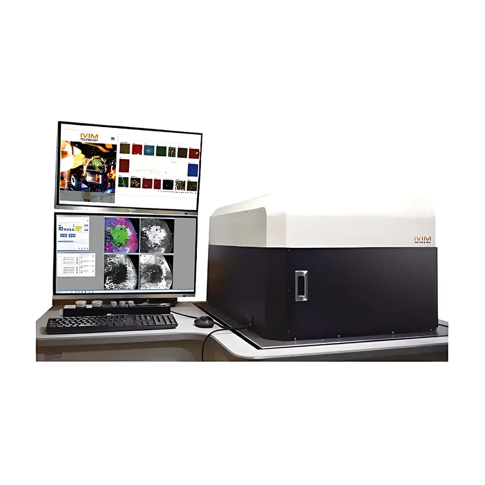
IVIM Small Animal In Vivo Confocal Imaging System
| Origin | South Korea |
|---|---|
| Manufacturer Type | Authorized Distributor |
| Origin Category | Imported Instrument |
| Model | IVIM |
| Price Range | USD 420,000 – 700,000 |
| Instrument Type | Optical In Vivo Imaging System |
| Energy Resolution | 4 keV |
| Scan Resolution | 512 × 512 pixels |
| Frame Rate | 100 fps |
| Field of View | 10 × 10 µm to 100 × 100 mm² |
| Sample Thickness Capacity | 4 mm |
Overview
The IVIM Small Animal In Vivo Confocal Imaging System is a high-performance optical imaging platform engineered for non-invasive, real-time cellular and subcellular visualization in live mammalian models. Based on point-scanning confocal microscopy principles, the system employs laser excitation and pinhole-confined detection to achieve optical sectioning with high axial resolution and minimal out-of-focus fluorescence background. Designed specifically for longitudinal studies in preclinical research, it enables dynamic monitoring of biological processes—including angiogenesis, immune cell trafficking, tumor progression, and metabolic activity—within intact, physiologically relevant tissue environments. The system integrates motorized XYZ stage control, multi-channel fluorescence detection (with spectral unmixing capability), and synchronized physiological monitoring interfaces (e.g., ECG, respiration gating) to support rigorous experimental reproducibility under controlled anesthesia and thermoregulation conditions.
Key Features
- High-speed confocal scanning at up to 100 frames per second (fps) with 512 × 512 pixel resolution, enabling capture of rapid physiological dynamics such as calcium transients or vascular perfusion.
- Wide-field adaptability via motorized zoom optics and macro-to-micro objective switching, supporting imaging across scales—from whole-organ surface mapping (100 × 100 mm² FOV) to subcellular detail (10 × 10 µm FOV).
- Dual-mode detection architecture: simultaneous spectral separation of up to four fluorophores using tunable emission filters and high-quantum-efficiency GaAsP photomultiplier tubes (PMTs).
- Integrated thermal regulation and stereotactic positioning system compatible with murine, rat, and juvenile rabbit models; supports head-fixed, dorsal, ventral, and lateral imaging configurations.
- Energy-resolved fluorescence detection with 4 keV energy resolution—optimized for distinguishing metal-chelating probes (e.g., Gd-based or Mn-enhanced contrast agents) and quantifying elemental co-localization in tissue sections.
- Rugged optical path design with active alignment stabilization and vibration-damped optical table integration to maintain focus fidelity during long-duration time-lapse acquisitions.
Sample Compatibility & Compliance
The IVIM system accommodates live small animal subjects with maximum sample thickness of 4 mm under standard imaging configuration—sufficient for superficial organ imaging (e.g., dorsal skinfold chamber, cranial window, ear pinna, cornea, or exposed intestinal loops). For deeper-tissue interrogation, optional near-infrared (NIR-II) excitation modules and adaptive optics correction units are available to extend effective penetration depth while preserving confocality. All hardware and software components comply with IEC 61000-6-3 (EMC emissions) and IEC 60601-1 (medical electrical equipment safety) standards. The system supports GLP-compliant workflows through audit-trail-enabled acquisition logs, user-access controls, and electronic signature readiness per FDA 21 CFR Part 11 requirements.
Software & Data Management
Acquisition, reconstruction, and analysis are managed via IVIM Control Suite v4.2—a modular, scriptable platform built on Qt/C++ with Python API extension support. Core functionalities include real-time Z-stack acquisition with autofocus feedback, multi-dimensional time-series registration (4D + λ), intensity normalization across sessions, and batch-processed deconvolution using measured PSF models. Export formats include OME-TIFF, NRRD, and HDF5 with embedded metadata compliant with the Bio-Formats specification. Integrated with OMERO server deployment options, the software facilitates centralized storage, role-based data access, and DICOM-SR export for cross-platform interoperability with PACS and preclinical imaging repositories.
Applications
The IVIM system serves diverse applications across oncology, immunology, neuroscience, and regenerative medicine. Validated use cases include: longitudinal tracking of GFP/RFP-labeled tumor xenografts in mammary fat pads; in vivo assessment of blood–brain barrier integrity using dextran-FITC leakage assays in cortical vasculature; real-time visualization of neutrophil extravasation in inflamed tracheal epithelium; functional mapping of retinal ganglion cell responses to patterned light stimuli; quantitative analysis of collagen fiber reorganization in healing dermal wounds; and spatial profiling of hypoxia-inducible factor (HIF-1α) expression dynamics in pancreatic islets under metabolic challenge. Its compatibility with standard transgenic reporter lines (e.g., CX3CR1-GFP, LysM-eGFP, Thy1-YFP) and commercial viability dyes (Calcein AM, Propidium Iodide) ensures seamless integration into established preclinical pipelines.
FAQ
What animal models are supported?
The system is validated for mice (C57BL/6, BALB/c, NSG), rats (Sprague-Dawley, Wistar), and juvenile New Zealand White rabbits. Custom stage adapters are available for zebrafish larvae and chick embryos.
Is anesthesia integration supported?
Yes—integrated ports for isoflurane vaporizer control, capnography, and pulse oximetry allow closed-loop physiological monitoring and automated acquisition triggering based on respiratory phase.
Can the system perform quantitative fluorescence intensity measurements?
Yes—calibrated PMT gain settings, reference standard slides (e.g., fluorescent microsphere arrays), and background-subtracted ROI analysis enable absolute intensity quantification traceable to NIST-traceable standards.
Does the software support third-party plugin development?
Yes—the Python API provides full access to acquisition parameters, raw detector data streams, and metadata structures, enabling custom algorithm integration for machine learning–based segmentation or motion correction.
What service and support options are available post-installation?
Standard coverage includes on-site installation and validation (IQ/OQ), annual preventive maintenance, remote diagnostics, and priority access to firmware updates and application scientist consultation. Extended SLA packages include 24/7 emergency response and dedicated training workshops.